Coomassie Densitometry and Loss
Previously I have silver stained all gels because the separations look very clear with this staining technique. However, the silver stain is not giving very quantifiable results. Coomassie staining means a loss in sensitivity, but it’s the gold standard for quantifying protein amounts. Here I’ve stained one of the protein separation results and rerun with coomassie staining.

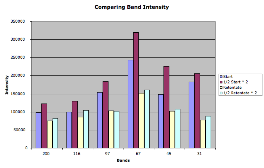
The bands are clear enough for densitometry, and first off I looked at how the intensity is related to the amount of protein. I took lane 2 and 4 (which have half the volume of sample) and doubled the intensity and compared to 1 and 3. In general this matches, but there is a good deal of error in the start solution especially.
I then measured the filtrate intensity and added these bands to the retentate. I compared the summation to the start sample, and this time it comes pretty close. Recall that in silver staining the retentates were usually the same intensity as the start sample and the summation with the filtrate was double the start sample. This was because the bands were saturated in the silver stained gels (I think). Now we can see a reduction in the retenate and a reasonable summation. The summation however tends to fall below the intensity of the start sample. This might be indicative of the loss of sample to the membrane/plastic.
