Rotation Project – Sergio Garcia
Introduction
The Ehler-Danlos Syndromes are a group of thirteen heritable connective tissue disorders. These disorders are genetic, and usually involve a mutation in genes that code for components in the collagen matrix (1). Some of the main characteristics in patients with these disorders include tissue fragility, joint hypermobility, and skin hyperextensibility (1). Classical EDS (cEDS), Hypermobile EDS (hEDS), and vascular EDS (vEDS) account for a vast majority of patients dealing with EDS (2). vEDS is the most mortal type of EDS as it negatively impacts the fragility of blood vessels and organs the most. People living with vEDS are more prone to aneurysms and rupture of arteries and organs. This occurs due to a genetic mutation in the collagen, type III, alpha 1 (COL3A1) which affects the synthesis of the pro-alpha (III) chain of collagen-III. Collagen-III plays an important role in the intima part of the vascular wall, between the endothelial cells and the smooth muscle cells. This is important given that collagen-III is a vital part of the structure of the vascular wall, and therefore its fragility. In addition to providing tensile strength, collagen-III is very important in wound healing as collagen-III is known to be a vital factor in the granulation of tissue after injury (3).
The original plan for my rotation project was based on this genetic disorder. I wanted to investigate the interaction between endothelial cells and smooth muscle cells on dual-scale micro+nanoporous membranes. However, given the limited time in the rotation as well as not having smooth muscle cells available to use, it was decided that the rotation project would consist of learning how to assemble the membranes, performing co-cultures with HUVECs on both sides of the membrane, and then co-culture of HUVECs-Pericytes.
Methodology
Human umbilical vein endothelial cells (HUVECs), pooled from many donors, were seeded on micro+nanoporous silicon nitride membrane chips (µSiM) on both sides of the membrane, creating a co-culture environment. Similarly, for the second model, HUVECs were placed on the top well and iPSC derived brain pericyte like cells were seeded on the bottom channel.
In both cases the devices were first coated with fibronectin. Media used for Pericytes was left on the top well, while the cells seeded in the bottom channel were placed. The device was flipped so the cells could settle at the bottom of the membrane interacting with the channel. After the cells were let to settle for a few hours, the device was flipped and HUVECs were seeded on the top side of the membrane.
The membranes used for the first model were fixed with formaldehyde and then stained with VE-Cadherin, which displays endothelial cell-cell junction integrity, and CD31 (also known as PECAM-1), a cell adhesion molecule that is highly expressed on the surface of endothelial cells. The second model followed a similar process as the first membrane. Cells were also fixed with formaldehyde and stained with VE-Cadherin, however, since Pericytes were part of this model, they were stained with platelet-derived growth factor receptor beta (PDGFR‐β antibody. Cells in both models were stained with Hoechst solution for nuclear staining. These devices were then imaged in a confocal microscope and analyzed using ImageJ-Fiij and Imaris Viewer.
Results
HUVECs-HUVECs Devices
VE-Cadherin and Hoechst staining are displayed in the image below. The membrane seems to have broken at the bottom right corner. However, it can be still be seen that there is formation of junctions between HUVECs. 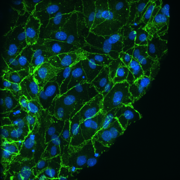
Similarly to the previous image, CD31 seems to be present in the image below, demonstrating the presence of HUVECs within the device.

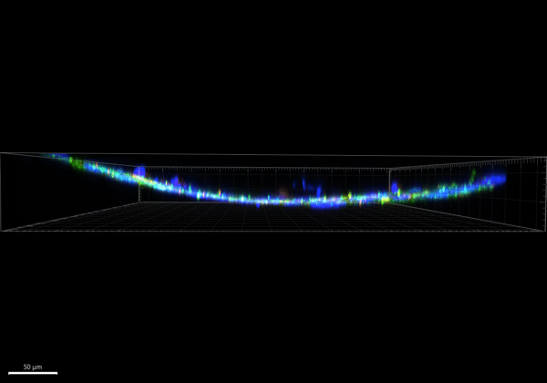
The images below display the 3D view of the dual-scale µSiM containing HUVECs on both sides. Based on these image and videos it can be seen that there are two layers of cells based on the Hoechst staining. The GIF video shown displays that there are cells at different z-positions. However, there given that the membrane appears to be broken it may be due to that that two layers are shown.

HUVECs-Pericytes Devices
The images below display Hoechst, VE-Cadherin, and PPDGFR‐β antibody staining. However, unlike the previous model, the staining seems to not stain the edges of the cells but rather the inside of the cells, for both VE-Cadherin and PPDGFR‐β. Hoechst staining seems to work fine.


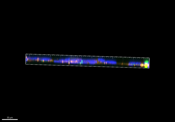
The image and video below display a 3-D view of the dual-scale µSiM. Unlike the previous model, this µSiM appears to not be broken and presents a nice view of the z-plane of the membrane.
Summary
The work done during my rotation in Dr. McGrath’s lab allowed me to get introduced to many different skills. Among the many of them the ones that I found to be most interesting was doing cell culture on these devices and working with the confocal microscope. As seen from the first model, the membrane appeared to be broken, but in later experiments as seen in the second model, my ability to work with these membranes got a bit better. Moreover, it can be seen from the first image that the staining worked on the first try. VE-cadherin and CD31, both markers for HUVECs were able to be seen, but on second model some of the staining appeared not to work as intended. However, while analyzing the 3D view of the membrane after staining, a nice view of the z-stack could be seen in the image and videos above. In the second model it could be seen that while there seems to be no markers for HUVECs present, the Hoechst staining displays some cells above the others, therefore revealing that there are cells present in both sides of the membrane. While this was my first try at both models, I believe that with more time, better images and therefore better results could be obtained. Overall, the work here has prepared me to at least have a better idea on how to work with these devices and to look out for certain steps if the work can be done with a vEDS model in the future.
Acknowledgements
I want to thank Julie Kuebel for getting me started on the assembly of these devices and teaching me a bit more on how to perform cell culture on them. I want to also thank Isabelle Linares for being able to help me with questions regarding the HUVECs used in my project. Lastly, I want to thank Michelle Trempel for helping me with the imaging and for helping me with the co-culture of HUVECs and pericytes as well as the staining of the cells used on these devices.
References
1. Royer, S. P., & Han, S. J. (2022). Mechanobiology in the Comorbidities of Ehlers Danlos Syndrome. Frontiers in cell and developmental biology, 10, 874840. https://doi.org/10.3389/fcell.2022.874840
2. Chiarelli, N., Ritelli, M., Zoppi, N., & Colombi, M. (2019). Cellular and Molecular Mechanisms in the Pathogenesis of Classical, Vascular, and Hypermobile Ehlers‒Danlos Syndromes. Genes, 10(8), 609. https://doi.org/10.3390/genes10080609
3. Volk, S. W., Wang, Y., Mauldin, E. A., Liechty, K. W., & Adams, S. L. (2011). Diminished type III collagen promotes myofibroblast differentiation and increases scar deposition in cutaneous wound healing. Cells, tissues, organs, 194(1), 25–37. https://doi.org/10.1159/000322399
Thanks Sergio. Close enough for a rotation! And one piece of inadvertent learning that helps illustrate for everyone that these membranes are flexible. They are only flat in practice because they are under tension on the chip. This can be appreciated from the piece that broke during staining which curls vs. the one that did not, which is flat.