Effectiveness of Nanosep Omega 300K centrifugal membranes in separating IgG conjugated qdots with excess free-floating antibody
Used Thermofisher SiteClick qdot antibody labeling kit 625. Began with 4.0mg/mL IgG and conjugated it with an unknown amount of qDots, methods provided by the SiteClick kit.
The final stage in the manual [1] is an optional cleaning of the qDots to remove any unconjugated and free-floating qDots from the mixture using the nanosep omega 300K centrifugal membrane. The manual reports an approximate 80% efficiency of the membrane in separating the free antibody. At this point we have been able to determine the overall amount of antibody flow-through and retentate, but one more calibration curve must be made in order to calculate the amount of free antibody cleared.
QDot Conjugation Stages:
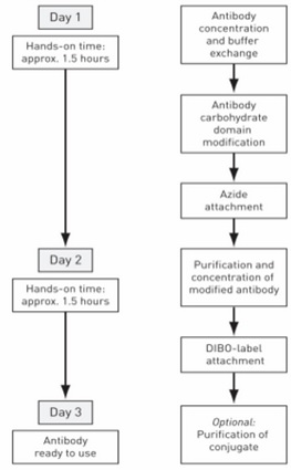 <– “Optional” stage is the target stage of interest
Measurement:
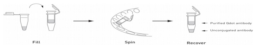
Collected 10uL sample of the initial 500uL volume of conjugated/free mixture. Then centrifuged the solution three separate times, each time re-diluting the pre-filtered solution so that each flow through was approximately 300uL. These flow-throughs were also collected for antibody content analysis afterwards. [1]
All solutions were tested at 280 nm absorbance for antibody concentration and zeroed at 250 based on calibrations Karl had done in earlier experiments (figure 1). They were also zeroed with water measurements in each cell.
Figure 1: Karl’s previous zero-ing at 250 using free IgG antibody and qDot-conjugated IgG

Table 1: Water Corrected Absorbance Measurements:
| 250nm | Cell 1 | Cell 2 | 280nm | Cell 1 | Cell 2 |
| initial | 1.81170 | 1.79620 | initial | 1.34090 | 1.30320 |
| I | 0.00460 | 0.00290 | I | 0.00270 | 0.00100 |
| II | 0.00440 | 0.00410 | II | 0.00320 | 0.00180 |
| III | -0.00130 | 0.00000 | III | -0.00170 | 0.00070 |
| final | 1.66020 | 1.64400 | final | 1.37580 | 1.28600 |
I-III: flow through antibody measurements
Figure 2: Absorbance fit curve for IgG
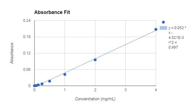
Water-corrected and A(280nm)-A(250nm) fit of the IgG antibody in tris based buffer solution
Based off the equation of the absorbance fit curve, antibody concentrations were found by using the 250-280 method:
(Absorbance + 0.004521)/0.052 = Concentration [mg/mL]
Table 2: Amounts of antibody at each stage
| [IgG] (mg/mL) | Cell 1 | Cell 2 | Volume (uL) | Mass (mg) | Cell 1 | Cell 2 |
| initial | 9.1408 | 9.5677 | 500 | initial | 4.5704 | 4.7839 |
| I | 0.1235 | 0.1235 | 262.4 | I | 0.0324 | 0.0324 |
| II | 0.1100 | 0.1312 | 334.2 | II | 0.0368 | 0.0438 |
| III | 0.0946 | 0.0735 | 353.6 | III | 0.0335 | 0.0260 |
| final | 5.5562 | 6.9716 | 318 | final | 1.7669 | 2.2170 |
| % | 60.78 | 72.87 | % reduction | 38.66 | 46.34 |
There is approximately a 42% mass reduction in the amount of antibody in the sample before and after separation with the nanosep omega 300K centrifugal membrane.
Qdot Fluorescence:
This week will make a qDot concentration fluorescence calibration curve so that we can back calculate the number of qDots in the initial and final mixtures. The numbers should be the same except for the 60-100uL spillage that occurred during the experiment (see below). Knowing the number of qDots will correlate to the assumption that there are 3 antibodies conjugated to each qDot, allowing the calculation of free antibody yield, instead of overall antibody yield.
Low flow-through readings (I, II, III) indicate that no qDots flow through the membrane.
Table 3: Fluorescence of qDots in initial and final volumes.
| Fluorescence | Cell 1 | Cell 2 |
| initial | 19409 | 19055 |
| I | 2 | 1 |
| II | 1 | 1 |
| III | 1 | 1 |
| final | 30456 | 29997 |
Things to consider:
- The main issue with this experiment is that after the first centrifugation stage (I), 60uL-100uL of the semi-purified qDot antibody was lost. The concentrations of either the qDots or antibody at this stage is unknown so the amount lost of either is unknown. The amount of lost antibody could be calculated if the concentration of collected antibody was high enough to have an absorbance reading. A potential solution to this is dehydrating the antibody flow-through so that most of the water evaporates and the antibody is left concentrated at the bottom. The initial volume is know and the final could be known by weighing the sample after the dehydration. The concentrated sample could then be used to get an absorbance value at 280 and 250 to know the amount of antibody filtered out from the initial centrifugation, so that the concentration of antibody in the lost 60uL-100uL could be determined and accounted for. The amount of qDot lost would be determined based on the initial amount of qDots and the volume it was lost at. The issue with this method is that the antibody may either stick to the sides of the container during dehydration instead of sinking to the bottom to concentrate, or the antibody may become denatured at the high evaporation temperatures so that it is no longer detectable.
- A second method is to use fluorescently tagged antibody in the next round so that their absorbance can be measured from the flow-through without manipulation.
- The two percent mass reduction readings above have a difference of 20% (in comparison to the 38.7% reading). These two measurements were taken from the same sample and were corrected using water correction and A(280)-A(250) correction. This large error indicates that further methods of standardization are necessary or the current method is flawed.
- Further analysis needs to be done in order to calculate the yield of free-antibody instead of total antibody. This will be done by doing a qDot fluorescence concentration calibration curve to determine the concentrations of qDots in the initial and final amounts. This will also be used to determine the amount of qDots lost in the process. The kit does not give the number or concentration of qDots provided, so all concentrations would have to be in reference to the unknown initially provided antibody amount.
The actual number of provided qDots must be determined in order to know how much antibody is free to be filtered out.
[[1] https://tools.thermofisher.com/content/sfs/manuals/mp10469.pdf1]