Cytotoxicity of silver chloride and platinum electrodes
I am working with different electrode materials as a substitute for ITO for my TEER measurement device. I used silver-silver chloride electrode and platinum-iridium ribbon (90:10) for that purpose. I made AgCl electrode by dipping about an inch of silver wire in silver chloride ink and letting it dry for half hour. I obtained the platinum ribbon from Waugh lab’s micropipette apparatus. I passaged cells (P4) and divided into three batches: control, silver electrode and platinum electrode. I did the imaging on day 4 and day 7. Results are self-explanatory!
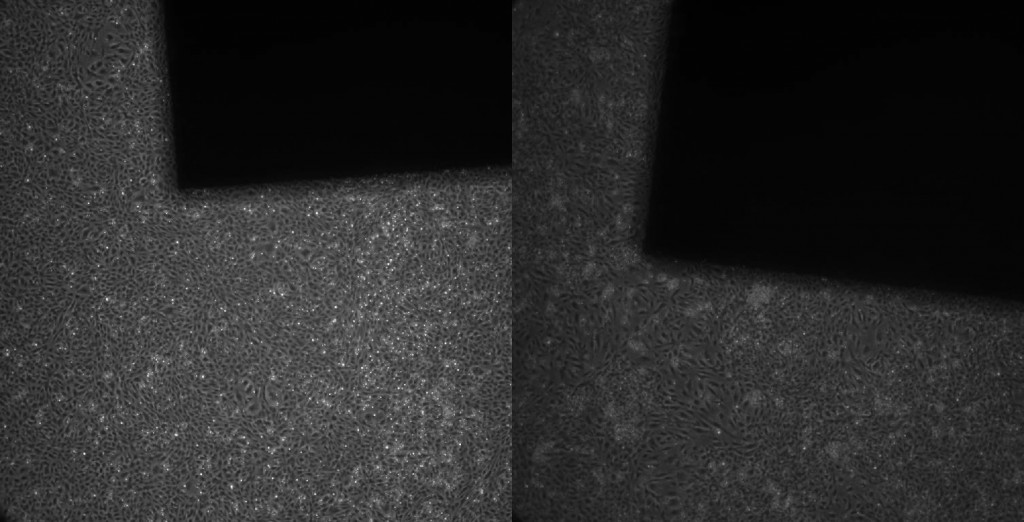
Control flask without any electrode, just cells and media

Flask with platinum electrode
Flask with silver chloride electrode

By day 7, the flask gets over confluent due to which there are many cells in field of view that are floating inside the flask. It can be seen in the control image, and also in the one with platinum electrode. Cells adjacent to silver chloride electrode are necrotic and dead. As we go away from this electrode, the viability of the cells increase, and at far extreme of the flask, cells display perfectly same morphology oblivious of the presence of the electrode. I will now be investigating about the usefulness of platinum as a material for my TEER device.