F-actin alignment on microporous SiO2 membranes
In our study of endothelial fibronectin fibrillogenesis and FA formation on microporous SiO2 membranes, we decided to investigate whether the pore orientation and pore spacing induced FA formation and therefore F-actin alignment. (Note: we updated our last post on FN fibrillogenesis here.) Spencer and I have been busy analyzing data and making figures.
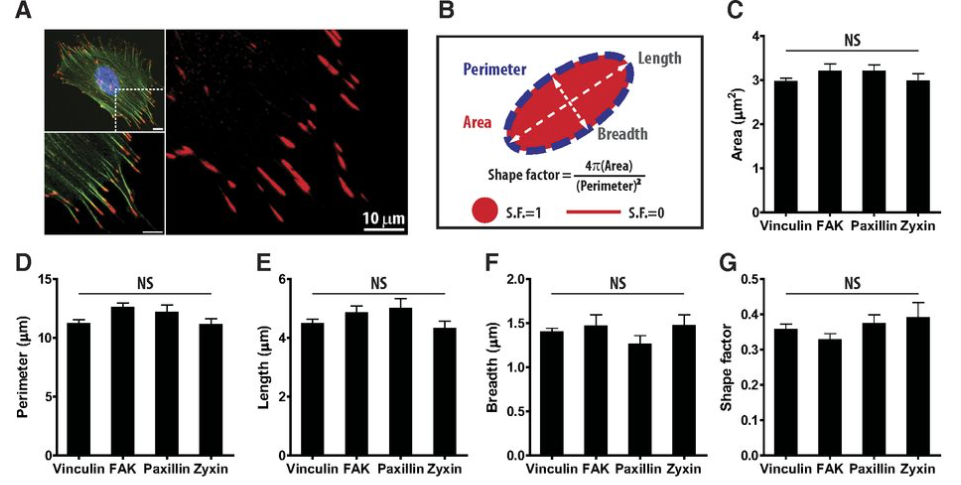
It has been reported that FAs have a defined size. That size varies by paper (and probably cell type/substrate), but it seems to hover around 1.5 x 4 microns. Here is a figure from Denis Wirtz that shows this with fibroblasts.
http://www.fasebj.org/content/27/4/1351.full
Based on the inter-pore spacing of our membranes, we hypothesized that FAs and their connection to F-actin would be aligned in a specific orientation. Since F-actin is aligned in the same orientation of the FA, we measured this since we felt our accuracy would be higher measuring F-actin fibers compared to smaller FA patches.
Since our 3.0 micron membranes have a nice “pocket” of contact area (30 degrees clockwise from vertical – see below), we expected to see F-actin predominantly in one direction on these membranes. We expected that non-porous membranes would have no preferred orientation. We also expected that the few FAs and F-actin that formed on 0.5 micron pore sizes membranes would be random and due to small contact areas and interactions with FN fibrils that overhung the small pores. We were wrong!
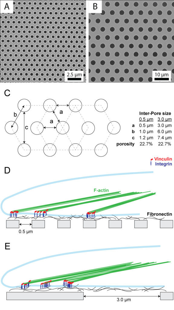
Note: Since hexagons have symmetry, we reported our measurements only within a 60 degree range. We mapped all results to this range and created the rose diagrams below. If you are as uncomfortable with geometry as I am, this may take you a while to appreciate.
As you can see from the figure below, there was relatively random F-actin orientation on the non-porous membranes as expected. The 3.0 micron pore size membrane also seemed to have somewhat random orientations, but possibly some clustering at specific angles. Interestingly, the cells on 0.5 micron pore size membranes did have a very strong preference for one single orientation. This alignment was not along the “infinite” track at 30 degrees from vertical (B), but rather at the vertical orientation (A). This may possibly be due to not only a length, but also a width requirement for the FA contact. See below for a graphical/geometrical explanation.
As a reminder: <20% of cells on 0.5 micron pore membranes formed focal adhesions (compared to ~100% on non-porous and TCP) and the cells formed <20 FAs (compared to ~50 FAs on non-porous and TCP). With this in mind, it is interested that these cells not only still formed FAs on regions of the membrane that are much smaller than the commonly reported FA size (and our own fluorescence images), but that the cells highly preferred an orientation that has a contact region of approximately 1 x 1.2 microns. This suggests that even though the cells are forming FAs on some FN that spans the pores, they prefer to form FAs on FN that is over the non-porous region of the membrane. Interestingly, the inter-pore spacing of the 3.0 micron pore membranes is large enough that the cells do not have a preferred orientation suggesting that the the size of the FN contact patch with the non-porous region of the membrane does not need to be as large as the fluorescence signal of the FAs (i.e. it can be as small as 3.0 microns, not 4 microns as reported by Wirtz).
