Unannealed MgF2 Tomogram
Previous work on Tomograms: TEM Tomography of NPN and npMgF2
Previous work on reconstructing the tomograms: Manual Reconstruction of Electron Tomograms
Introduction
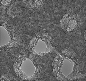
This post examines another set of MgF2 pores that Kilean acquired. I want to compare the inner pore morphology of two different processing flows for MgF2 nanomembranes, one of which gives us excellent SERS properties (250C heated evaporation) and one that does not (Room Temp evaporation, Annealed at 600C). I suspect that the internal pore roughness of the annealed films are much smoother than the heated deposition. Increasing surface roughness has been shown to increase the SERS effect tremendously, and creating even rougher characteristics may improve the behavior of our devices.
Dataset
Reconstruction Settings
I used TomoJ (which is now installed on the Mcgrath lab mac pro in Fiji) to perform the reconstructions
- Dataset rescaled to ~400×400 pixels (originally 2048×2048)
- Dataset Aligned using SIFT (Scale Invariant Feature Transform) registration in Fiji
- Default settings
- Dataset cleared of hotspots
- 1 pixel radius
- Dataset background subtraction
- 100 pixel rolling ball
- Aligned with Crosscorrelation feature
- Default settings
- SIRT (Simultaneous Iterative Reconstruction Technique) Algorithm
- 100 iterations
- Diminishing returns beyond 30, but still some improvement
- 1.0 relaxation coefficient
- resin/cryo sample
- 400 pixels thick
- About 1hr reconstruction time (400x400x400 voxel array)
- 100 iterations

Once the tomogram had been created, I manually traced out some contours using FreeD, which were then projected into a 3D space.
Discussion
- There are a few alignment errors in the original tilted image; which translate to blurriness and noise in the reconstructed tomograms. A potential way to improve this stack is to use something like 3D alignment, instead of the crosscorrelation alignment algorithm that I am using now
- The raw data set shows the interior surfaces of the nanovolcanoes as having rough granules of MgF2 throughout the surface. My tomogram captures this feature as contours are jagged throughout the volume stack. However, I did not trace out individual contours to highlight this feature in the FreeD surface projection. I am still working out the best method to automate the contour segmentation which will give us another of confidence.
- I had to split up the bicavity into 3 different surfaces to have it display properly in FreeD. The orange structures represent the inner part of the nanovolcano, and where it bifurcates, I made the new contour red.
- The grains of MgF2 (pink) are easier to pick out in the surface plot. We can identify a front and back surface readily.
- Some of the sidewalls of the cones seem to be a lot smoother than what the MgF2 nuggets appear like in the bulk film. Is it a different material (contamination)?
- There’s more work to be done on processing contours automatically instead of relying on my hand drawn interpretation; this should better reveal jaggedness on the inner surface of the tomograms.