MgF2 cytocompatibility #3
I have been trying to verify that my MgF2 material is cytocompatible (e.g. no different than tissue culture plastic), and attempted Live/Dead staining to confirm. The following images are from P6 HUVECs with Invitrogen L3224 Live/Dead stain on MgF2, NPN, and tissue culture plastic after ~96 hours. The devices were initially seeded at 5e5 cells/mL, and the membranes were isolated with 300 micron gaskets from each surface. Fixation was accomplished with 3.7% formaldehyde, and permeabilized with 0.1% triton.
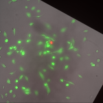
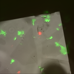
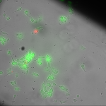
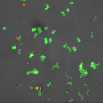
The MgF2 fractured after repeated wettings, but showed the same proportion of live and dead cells.
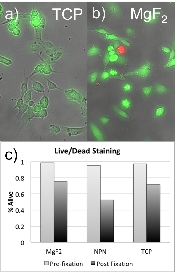
There were 40-80 cells in each of these measurements. However, the figure is only n=1, and I need to repeat the experiment to gain some estimate of error. I only have 1 large area freestanding MgF2 nanomembrane left, but I am in the process of making some more MgF2 nanomembranes with smaller areas. I think I have finally demonstrated similar cell morphology and survival outcomes across substrate types with this experiment.