Bend.3 + Geltrex Tube Formation Studies
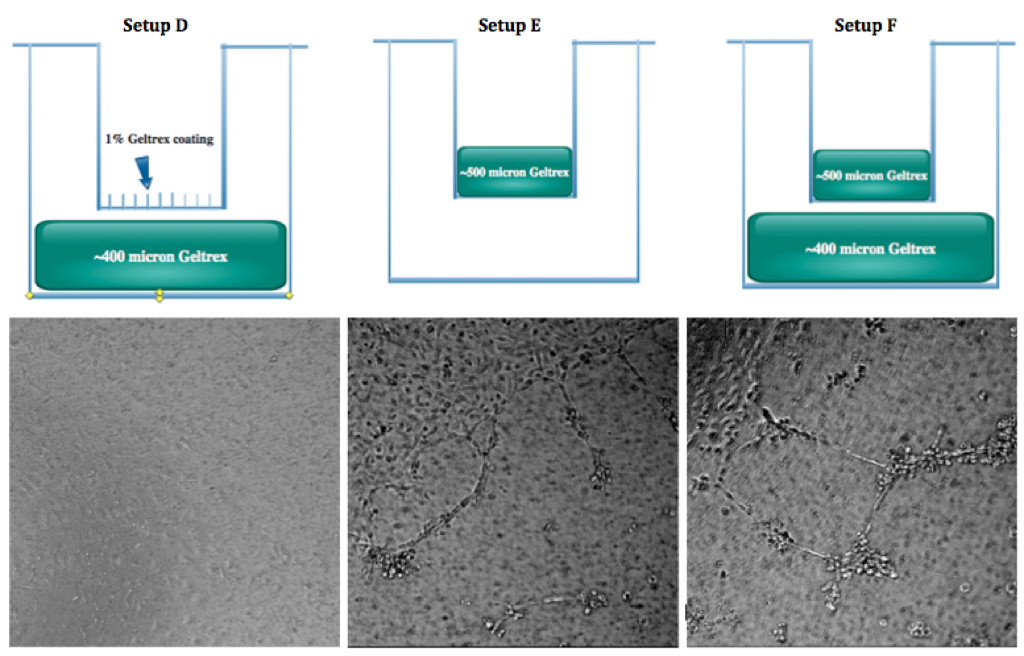
The following diagram lays out each separate setup that was tested using different combinations of Geltrex and substrates. Each trial has a different reasoning behind why it was done. The purpose of these trials was to investigate the effects of Gelrex gel and PCTE membrane inserts on tube formation in Bend.3 cells.
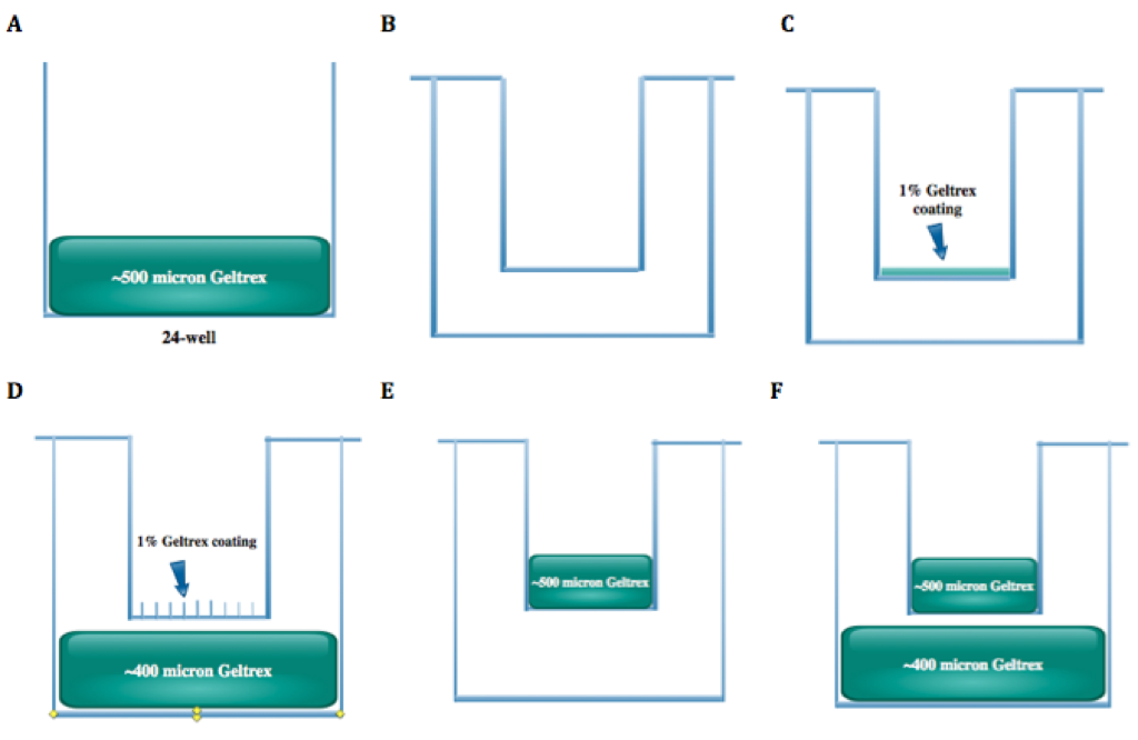
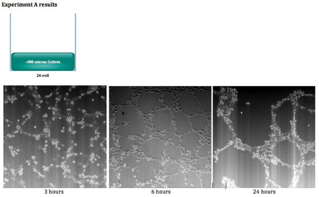
First, we showed that Bend.3 cells will successfully form tubes inside a 3D gel of Geltrex. We saw rapid tube formation in a 24-well with a thick gel (approx. 500 μm thick) where no membrane was present (setup A).
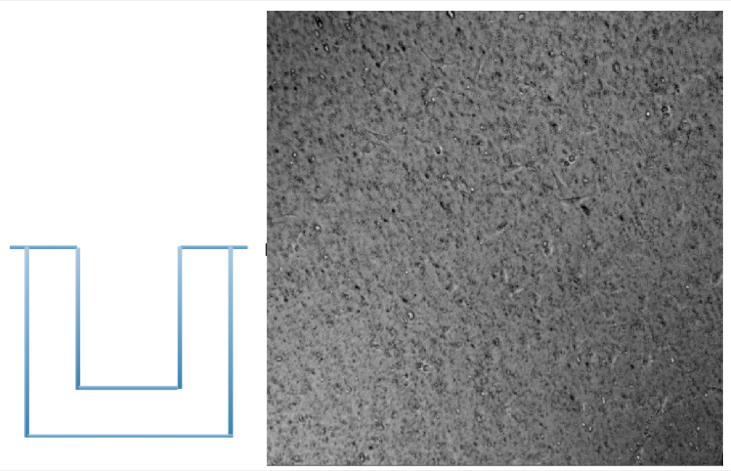
Bend.3 cells were then grown on 0.4 μm membrane inserts inside a 24-well and surrounded by media on the top and bottom to form experiment (B). There was no coating of Geltrex applied. This was to investigate if any tubes would form simply by the addition of a membrane alone. The cells adhered and spread out happily, but did not form any significant tubes.
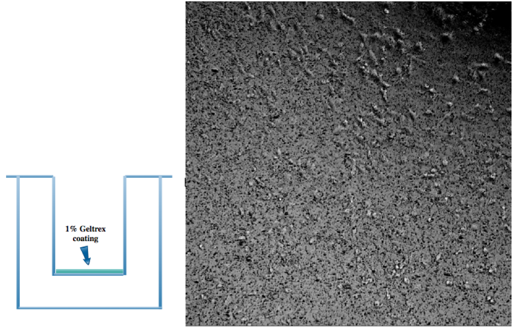
Next in experiment (C), we tried coating the inserts with a 1% Geltrex coating to provide proteins for the cells to adhere to and to test whether this would improve tube formation. The cells seemed to spread out more, but again, no true network or tube formation was seen.
Therefore, we wanted to combine the thick gel that did form tubes with membranes and discover wether the membrane was preventing the tube formation or simply not encouraging it when implemented alone. One setup (D) tested cells growing in a membrane insert inside a 24-well with a thick Geltrex gel underneath. The gel thickness was reduced to 400 μm to avoid the gel touching the bottom of the membrane and potentially cells. This was to test whether any growth factor inside the gel would leach out and facilitate any tube formation. The 2nd setup (E) called for a thick Geltrex gel inside a membrane insert with cells seeded inside, and media on the outside. This mimicked the setup of B while also adding a membrane. Finally, the 3rd setup (F) had the 500 μm gel inside the insert and also at the bottom of the 24-well (400 μm thick). (F) increased the total volume of Geltrex and therefore the growth factor concentration in the system.
After 24 hours, Setup A saw little to notube formation similar to the 1% Geltrex coating. Setup B’ saw small isolated tubes and slow formation, but they never managed to form an intricate network like the plain gel alone did. Setup C saw the best tube formation of the three. It had small monolayer areas but tubes extended from these and met up with other neighboring tubes to form small networks. It also had few areas with tubes growing in different planes. This suggests that the added growth factor did help the cells along the process.
Finally, we added a trial (G) where the thick gel in experiment (A) would be allowed to soak in media for 45 minutes and changed out three times to effectively dilute the amount of growth factor present in the system. This could then be compared to the previous results to determine if less growth factor but the same 3D environment would decrease tube growth.
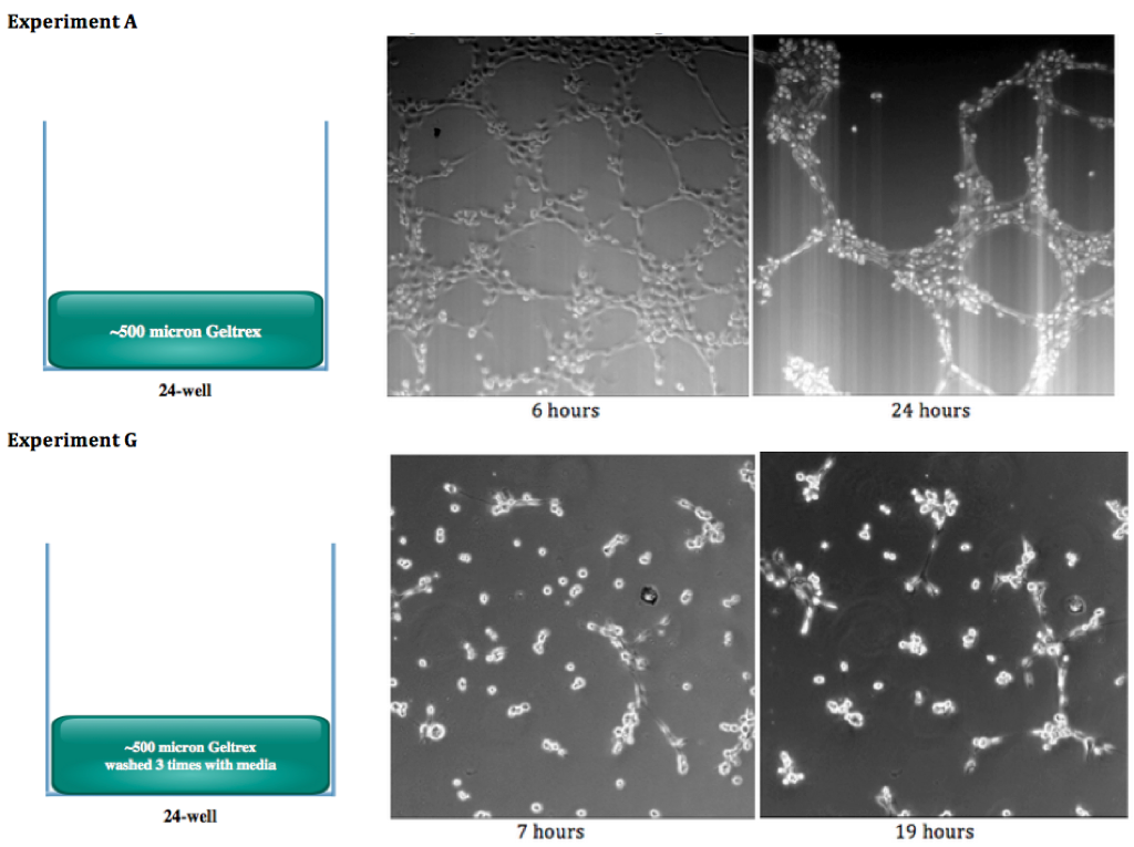
There were significantly less tubes, and thecells tended to clump togetherlike they do early on in the process where Geltrex growth factor is not diluted. This suggests that the 3Denvironment of a porous gel alone will not produce tubes as well as the same setup with growth factor. This also implies that growth factor is potentially more important than the physical/spatial clues of a matrix. We are working on moving to the same type of testing with HUVECs, with collagen gels instead of Geltrex, and using CytoVu chips instead of the PCTE membrane inserts.