Scanning ion conductance microscopy of cells on TEM grids (pnc-Si, not annealed)
During the recent visit of Greg Madeijski to our Institute of Biophysics, Imaging & Optical Science (IBIOS) here in Nottingham we had the opportunity to try imaging cultures of cells on several nanoporous membranes, including CytoVu’s and also the unsupported TEM grids without SiN.
Here is a short summary of our adventures, along with some encouraging preliminary pictures and some background on the scanning ion conductance microscopy (SICM) technique.
SICM is similar to atomic force and other scanning probe microscopies in that a tip is used to profile the surface of samples to build very high resolution images. An important advantage of the method is that it is highly optimised for biological samples as it requires the presence of conductive bathing media to work. In SICM a glass capillary-based microelectrode (similar to a patch clamp pipette) is used as the probe, which is moved in XYZ in a tapping mode to build the image:
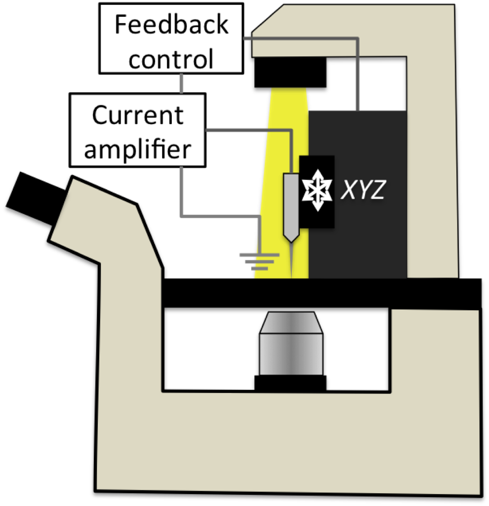
Feedback to detect the surface is supplied by passing a constant current from the electrode tip into the bath, and when the pathway for conduction is impinged upon by approaching the surface the electrode detects a change in current via a sensitive lock-in technique prior to contact. The amount of feedback controls the current decrement at the electrode required to trigger the feedback. Maximum resolution is dictated by the internal radius of the electrode tip, which can be down to several nanometers. There are the usual tradeoffs to be made between speed and sensitivity – control of the electrode is limited ultimately by the inertia of the scanning aparatus, and has been compared to flying a jumbo jet across bumpy terrain while keeping only a few centimeters above the ground!
We have had some trouble keeping cells happy and stuck to the CytoVu membranes, which we’re presently trying to optimise. Taking a membrane on which cells have been growing but floated away, we can see some evidence of residue either of the coating we applied before the cells, or secreted by the cells themselves:
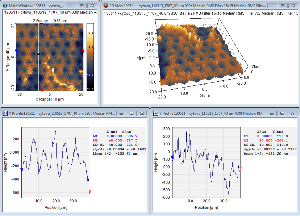
Overall vertical range is 1.94um – accounting for debris visible lower right. Sinusoidal appearance of SiO surface draped over SiN support – this is imaged apical side up, after cells were on the membrane for approx 5 days. Pre-coating was Poly-L-Lysine in this case, and the cells were ARPE-19 (human retinal pigment epithelium).
Looking at an 8um CytoVu, SiN side up:

You can see here that the 8um features are nicely profiled by the instrument. There is some striping due to thermal drift during scanning, which could be removed but which has not been processed here. The vertical scale is ~1um, only to account for some dust or something which appears as lightish blobs on the left side of the image. Field of view is in this case 40x40um (max possible 98um)
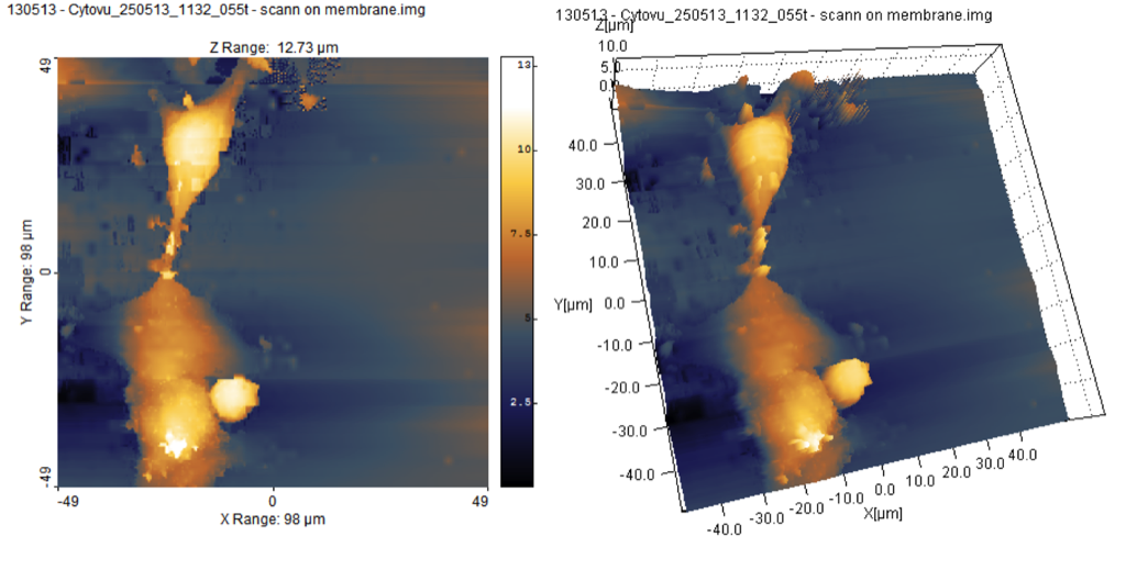
Zooming in with a subscan as shown (12.7×12.7um) we can map a single micropore:

In this measurement we read closer to 200nm for the depth – there is again some sloping due to thermal drift (in process of being solved by boxing the rig)
We also had a look at some fixed cells on CytoVus, in a case where the cell adhesion was better. The following images demonstrate some issues that can happen when scanning preps where the cells are a little unhappy:
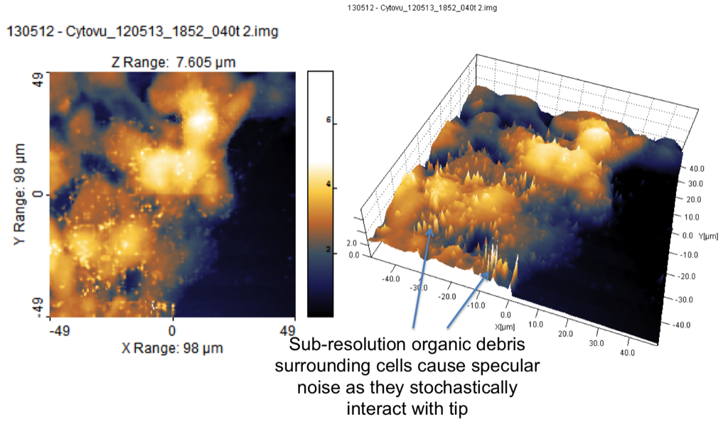
Debris can block the tip – either transiently or permanently – and lead to noisy images (spikes show where feedback has been inappropriately triggered far from the cells by debris floating in the bath or hanging around the fixed cells).
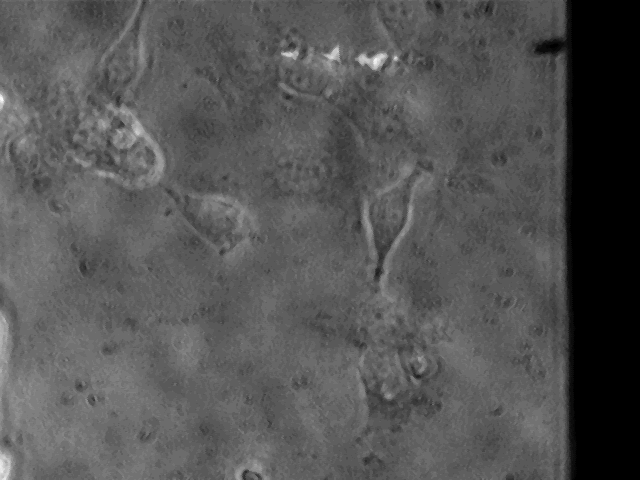
Greg and I played a little with imaging some cheek cells (buccal epithelia – just scrape the inside of your cheek gently and you’ll always have a sample to test your imaging equipment!).
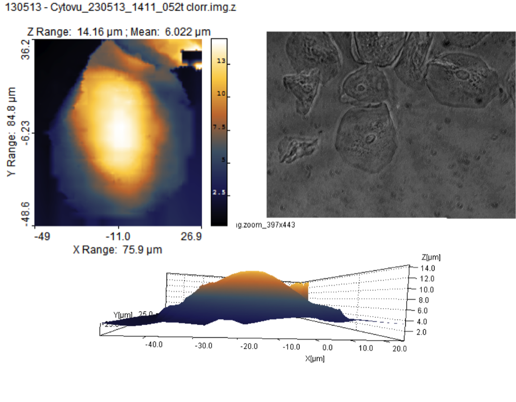
You can see here the excellent SNRwe can achieve in this instrument, and also a phase contrast view of the same cell taken using my novel condenserless phase contrast imaging setup (on which more another time).
Back to the pnc-Si: we looked at some cells which had been plated on some un-annealed TEM windows kindly sent over from Simpore. These unsupported membranes improved the phase contrast imaging of the cells, but heavily attentuated fluorescence signals in the UV – we need to look at the optical properties of these membranes or to turn the cells over for imaging. Obviously that would mean no SICM as the electrode approaches from the top.

- cells on non-annealed TEM grid. Red: CellMask, Cyan: Hoechst
Imaging on these substrates we noticed something interesting. I am accustomed to using feedback criteria of the order of 0.5% current decrement to image cells, but using this setting resulted in the electrode punching straight through the pnc-Si membrane as if it wasnt’ there. It didn’t seem to damage the probe to do this, and we might be able to do this deliberately in future to make small defects or slits in the membrane for control measurements. Greg and I had the feeling that the membrane was so permeable to current that perhaps it wasn’t actually interrupted the ion flow enough to give a feedback signal. Scanning near some cells on the edge of the membrane we noticed that 0.5% feedback in hopping mode was actually causing “trampolining” (coined the phrase to describe the below):
Dialling back the feedback to 0.2% (requiring careful setup of electronics!) allowed me to obtain the image below of the same cell, without any visible trampolining and a reasonable SNR:
The images aren’t too bad! Overall height is just over 12um, and the cells are not noticably blebbing or looking terribly stressed. There are however apoptotic remnants in the prep (small ball lower left).
This image was being acquired overnight as Greg left Nottingham, and took nearly 90 minutes to acquire as I had to s l o o w w w everything right down to accomodate the low feedback signal. However, some early success!
Here are some further tests I have made since, demonstrating an enhanced fluorescence detection (by upping the dye concentration unreasonably high) and using a faster scan protocol with 0.2% feedback. This was possible by targetting cells very close to the edge of the unsupported pnc-SI membrane, where I thought flexion and instability would be minimised:

Here again there are some apoptotic bodies around, and the cell to the top right of the scan looks a little blebby to me. I can, however, see the shape of some Nature figures at some point 😉
To really challenge the instrument and my own patience I tried to image the membrane again after a further 48hrs in media (cells were fixed so stayed around).
I found that the non-annealed pnc-Si was very fragile indeed by this point and showed several spontaneous tears and other defects in places even where we hadn’t been hammering it.
I figured that cells lying near a tear would be the most unstable subjects possible, since the membrane would be more or less completely unsupported, and has obviously become even thinner over time:

Now I wouldn’t call this a complete success, and the image again took nearly 100 mins to acquire, however before the electrode became blocked by debris in the prep I was able to image the cells even right up to the edge of the tear(!) Cropping out the dud signal from the deteriorating electrode, we are left with a relatively interesting reconstruction:
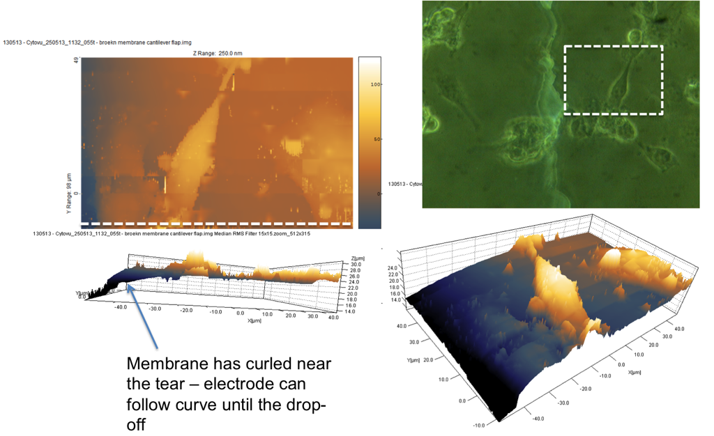
This data has been cropped but not processed, and shows the actual morphology of the cells as well as the curling of the pnc-Si substrate as the electrode approaches the tear. It is possible this curvature represents flexing of the membrane rather than an intrinsic curve, but it is very interesting nonetheless! The electrode has faithfully tracked 10um of membrane deflection, whilst imaging morphology on a patently unstable and ruptured pnc-Si substrate. This is very promising for future measurements as I should be able to look at morphological dynamics of cells on these substrates over time – perhaps looking at nanoscale features like filopodia and lamellopodia as the cells establish and come to confluence.
Scientifically, this is where I leave you, but we are looking forward to having Greg here again soon to crack on with making some devices to allow confluent epithelial cultures on these unsupported membranes, which we will be using for transport assays in parallel with some advanced imaging. SICM is only part of the story to come, but I hope you found this interesting nonetheless!
As well as geek stuff, here is Greg with Dr Emilia Moradi (our cell culture guru) and myself at an excellent Iranian restaurant in Nottingham called “Debsh” (translates as something like “Yum!”) :
Iranian food is awesome. we had Keskh Badenjoon (eggplant dip with garlic and chicken – my favouriate!) and Koobideh (traditional kebabs, done in a special way on long swords over a charcoal fire). Food is served with rice flavoured with saffron, which gives it a lovely buttery colour.
I look forward to meeting the group in September, when I will be visiting Rochester under a HERMES travel Fellowship to discuss microfluidic devices for electrophysiology and imaging of transporting epithelial systems.
All the best
Kevin
“““““““`
Kevin F. Webb, PhD
Royal Academy of Engineering/EPSRC Senior Research Fellow
Institute of Biophysics, Imaging & Optical Science
School of Electrical & Electronic Engineering
University of Nottingham
Phone/Fax +44 115 846 6580
kevin.webb@nottingham.ac.uk
I did some research on the blog, Jess Snyder made a nice post here about the transmission of the pnc-Si
https://trace-bmps.org/data/2008/03/05/transmission-spectra-of-pnc-si/
https://trace-bmps.org/uploaded_images/Picture-5-771254.png