
IMR90 EECM-BMEC-Like Cells Lack P-gp Efflux Activity
Introduction
During the January TraCe-bMPS progress meeting, Jon Flax presented some interesting findings in the iPSC-derived extended endothelial culture method brain microvascular endothelial cell-like cells (EECM-BMECs, hereafter referred to as BMECs) [1]. While these BMECs tended to have high expression of most transmembrane transport proteins, they had relatively low expression of p-glycoprotein (ABCB1/MDR1) (Figure 1).

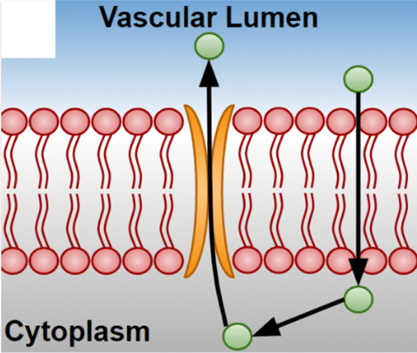
P-glycoprotein is an active efflux transport protein that is highly expressed on the luminal surface of brain endothelial cells in vivo. When lipophilic molecules diffuse through the endothelial plasma membrane, they are pumped back out to the blood via p-glycoprotein or other efflux transporters such as Breast Cancer Resistance Protein [2] (Figure 2). This pumping activity contributes to maintaining the blood-brain barrier. It also provides a challenge for drug delivery to the brain; a lipophilic drug may be able to cross through the luminal endothelial plasma membrane, but may fail to cross the abluminal plasma membrane into the brain parenchyma if it is pumped out of the cell by p-glycoprotein.
Because of their low p-glycoprotein expression as measured by RNAseq, it is unclear whether the BMECs will be useful for certain drug transport studies. To investigate the effective pump activity, we treated BMECs with rhodamine-123, a fluorescent substrate of p-glycoprotein.
Methods
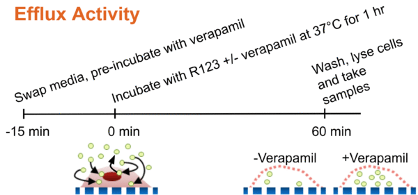
BMECs or hCMEC/D3s were seeded in 96-well plates coated with 10 µg/ml collagen IV or 100 µg/ml collagen I, respectively. 7 – 8 days after seeding, the cells were pre-treated for 15 min with 50 µM verapamil, a calcium-channel blocker known to inhibit p-glycoprotein activity, or a vehicle control. Following the pre-treatment, 10 µg/ml rhodamine-123 was administered to the cells with additional verapamil or vehicle control. After incubating for 1 hr, the cells were washed with PBS to remove any external rhodamine-123 and lysed with RIPA buffer (Figure 3). Fluorescence intensity of rhodamine-123 was then measured on a plate reader with the excitation wavelength set to 485 nm and emission wavelength set to 525 nm. Simply put, this was a measurement of all the rhodamine-123 dye that was still located inside the endothelial cells after the washing step. Higher pump activity would manifest as a lower fluorescence reading, while pump inhibition with verapamil would lead to a higher fluorescence reading.
Results
We first ran the experiment with the BMECs and found that there was no difference in rhodamine-123 fluorescence intensity with or without verapamil treatment (Figure 4). This could indicate two possibilities: either the verapamil was ineffective (it is possible that it had undergone too many freeze-thaw cycles or that it had expired) or the BMECs did not have any effective pump activity at baseline. To examine whether the verapamil was working properly, we repeated the experiment with the hCMEC/D3 cell line, which we had previously used to validate this assay. The hCMEC/D3s demonstrated a significant increase in rhodamine-123 fluorescence signal with verapamil treatment. This confirmed that the verapamil was working properly.

Note that the measured rhodamine-123 fluorescence intensities differed greatly between the two experiments despite the fact that the same concentration of rhodamine-123 was used in both instances. There are a few possibilities that may have contributed to this difference. Although both experiments used the same concentration of rhodamine-123, it is possible that the stock solution was not properly mixed before each experiment. The BMEC data also was subjected to background subtraction using a “blank” well (a mix of RIPA buffer and PBS), but there appeared to be rhodamine-123 contamination in the blanks for the hCMEC/D3 experiment, so this subtraction was skipped.
Conclusions
It appears that the BMECs do not have functional p-glycoprotein, which matches the low p-glycoprotein expression levels as previously determined by RNAseq. These results were obtained with BMECs derived from IMR90 cells. It should be confirmed whether the low p-glycoprotein expression is a result of the differentiation method or whether this is exclusive to the IMR90s, especially since other iPSC sources (e.g. SCTi003) will be used for the TraCe project.
References

Discussion summary:
Jon Flax noted that P-gp is also not expressed when other cell lines are derived using the EECM-BMEC protocol. I believe as many as six lines have been tried and all are low in P-gp expression. I updated the post with Jon’s slides from January. So this is not going to be remedied by changing donors. Might also be a maturation issue with iPSC-derived cells which is a common problem.
We agreed that engineering P-gp over-expression into a iPSC line may be a project worth undertaking. We will turn our attention to this idea next.
There are also a number of other channels that are expressed successfully (see Jon’s slides). Testing the function of these should also be on the docket. Because of some overlap between efflux transporters, having a non P-gp expressing line could be ‘feature’ instead of a ‘bug’ if there is interest in these specifically (credit Ben Miller).
Louis – Please take a minute to update your cartoon so that it shows expectations for functional vs. blocked P-gp function.
Hi Jim,
The efflux assay cartoon has been updated per your request.