Vascularizing the Retinal-Barrier-on-a-chip Part 1: Key Factors in Preparing the Fibrin Gel
By Rufaro R. Gamariel, Kevin Ling, James McGrath
Introduction
The retina enables visual perception by converting light to electrical signals, which are sent to the brain (Nguyen et al., 2023). It comprises two types of photoreceptors: cones are responsible for high acuity color vision, and rods are sensitive in low light conditions. The outer blood-retina barrier (oBRB) supports photoreceptors by regulating nutrient/ion transport and phagocytosing photoreceptor outer segments. The macula is a specialized region located at the center of the retina near the back of the eye that is densely packed with cones. It is responsible for providing crisp, detailed vision. Age-related macular degeneration (AMD) originates in the oBRB, which comprises retinal pigment epithelium (RPE) cells, Bruch’s membrane and the choriocapillaris (Song et al., 2023) as shown in Fig. 1A. The choriocapillaris is important because it supplies oxygen and nutrients to the outer retina (Lejoyeux et al., 2022). Disruption of the oBRB causes the death of RPE cells and photoreceptors, ultimately resulting in the loss of central vision (Abcouwer et al., 2024).
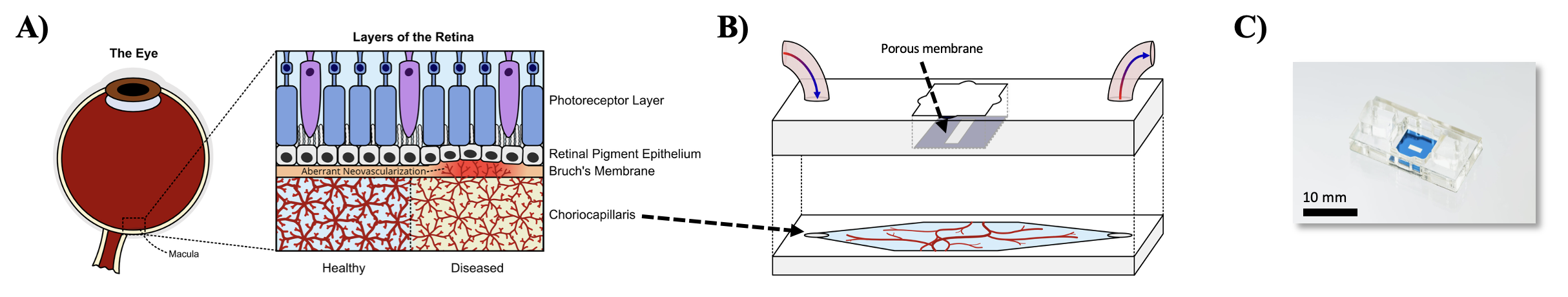
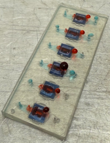
Figure 1: The μSiM-oBRB device mimics the layers of the retina in vitro. A) The retina comprises the choriocapillaris, Bruch’s membrane, an RPE layer and a layer of photoreceptors. In disease, blood vessels from the choriocapillaris penetrate through Bruch’s membrane towards the RPE monolayer. B) A vascularized hydrogel can be developed in the basal channel of the μSiM-oBRB device, a porous membrane separates the apical and basal chambers and an RPE monolayer can be cultured on top of the porous membrane. C) Photograph of the fully assembled μSiM-oBRB device.
Current drug development for AMD focuses on therapies that target inflammation, abnormal blood vessel growth and retinal cell degeneration (Rodriguez-Cruz et al., 2025). However, drugs often fail before they reach the market because of failures in safety testing, and even fewer show efficacy. Animal models such as mice lack maculae, and monkeys, despite being similar to humans and having the ability to develop AMD, grow and reproduce very slowly. Herein, we describe a protocol for developing a vascularized retina-on-a-chip platform (μSiM-oBRB chip; Fig. 1B–C) featuring a monolayer of adult RPE cells (ARPE-19, ATCC), which is physically separated from a fibrin-based hydrogel containing a co-culture of human umbilical vein endothelial cells (HUVECs, Vec Technologies) and human mesenchymal stem cells (hMSCs, derived from a donor). The apical well of the μSiM-oBRB was separated from the basal chamber using different porous membranes: 0.5 µm diameter microporous membranes, nanoporous membranes, and dual scale membranes which have both nanopores and larger patterned micropores (3 µm diameter) (SimPore, Inc.). We investigated whether usage of different membrane formats affects 3D vasculogenesis in the bottom compartment of μSiM microvascular networks. We hypothesized that microporous and dual-scale membranes would promote the generation of denser microvascular networks with higher average vessel diameter. We selected HUVECs for their robust angiogenic potential (van Duinen et al., 2020) and hMSCs for their supportive role in vessel maturation and stability (Summer et al., 2022). Collectively, these cell types spontaneously generate capillary-like vascular networks when co-encapsulated in supportive 3D ECM hydrogel matrices (Kurokawa et al., 2017; Shirure et al., 2021; Shirure et al., 2017). Fibrin was chosen because it is known to support endothelial cell migration, tubulogenesis and anastomosis in microphysiological systems (Moura et al., 2024; Shirure et al., 2021). Fibrin is formed when thrombin is mixed with fibrinogen; thrombin is a catalyst that converts soluble fibrinogen into insoluble fibrin by cleaving small peptides allowing the fibrin molecules to polymerize into a matrix (Jarvis et al., 2003; Shirure et al., 2017).
Further, we also hypothesized that apical vascular endothelial growth factor (VEGF) stimulation would induce migration of HUVEC vessels into the apical ARPE-19 chamber, simulating choroidal neovascularization as seen in wet AMD disease progression.
Methods
The μSiM-oBRB chips were assembled on Day 0. To sterilize the devices, all components except the membranes were soaked in 70% ethanol and allowed to dry before assembly. The assembled devices were further sterilized under UV light for 15 minutes and stored in sterile conditions. The devices were primed by adding 100 μl of Dulbecco’s Phosphate-Buffered Saline (DPBS; without calcium and magnesium ions) into the well and basal channel of each device. 10 mg/ml fibrinogen (from a 25 mg/ml stock) was prepared in DPBS (pH ~7.4) 1 day before cell seeding, sealed with several layers of Parafilm, and incubated in a 37 ℃ water bath overnight. PBS (pH ~7.2) was also used to prepare some of the fibrinogen. The devices were primed a second time using Vasculife endothelial cell growth media (Lifeline Cell Technology). We prepared fibrin-based hydrogels using previously described methods (Kurokawa et al., 2017; Shirure et al., 2021; Shirure et al., 2017). The fibrinogen was centrifuged at 200g for 2 minutes and filtered using a 0.22 μm low protein binding syringe filter.
Next, thrombin (from a 100 U/ml stock) was diluted with DPBS to a working concentration of 4 U/ml. For each device to be seeded, an individual fibrin-cell suspension was prepared by resuspending the desired number of cells (3 × 10⁶ or 5 × 10⁶ cells/ml at a 2:1 ratio of HUVECs to hMSCs) in thrombin (4 U/mL), followed by the addition of fibrinogen (10 mg/mL) to achieve final thrombin and fibrinogen concentrations of 2 U/mL and 5 mg/ml respectively. 20 μl of this fibrin-cell suspension was quickly injected into each channel. Care was taken to not to introduce bubbles into the basal hydrogel. The seeded devices were incubated for 30 minutes (37 ℃ and 5% CO2), then 100 μl of endothelial cell media was added to the well. μSiM devices were maintained on a 150 mm diameter petri dish during cell culture. A reservoir filled with DPBS was included to maintain a humid environment. Media changes were performed every two days, and the devices were maintained for up to 14 days in a standard incubator.
The samples were fixed in 100 μl of 4% paraformaldehyde for 20 minutes and washed with an equal amount of DPBS on Day 14. They were permeabilized and blocked for 1 hour using a buffer comprising 1% bovine serum albumin, and 0.25% Triton X100 mixed in DPBS. The samples were stained via immunohistochemistry for confocal imaging. The samples were imaged using the Andor Dragonfly Spinning Disk Confocal Microscope.
Results and Discussion![]()
![]()
![]()
![]()
![]()
![]()
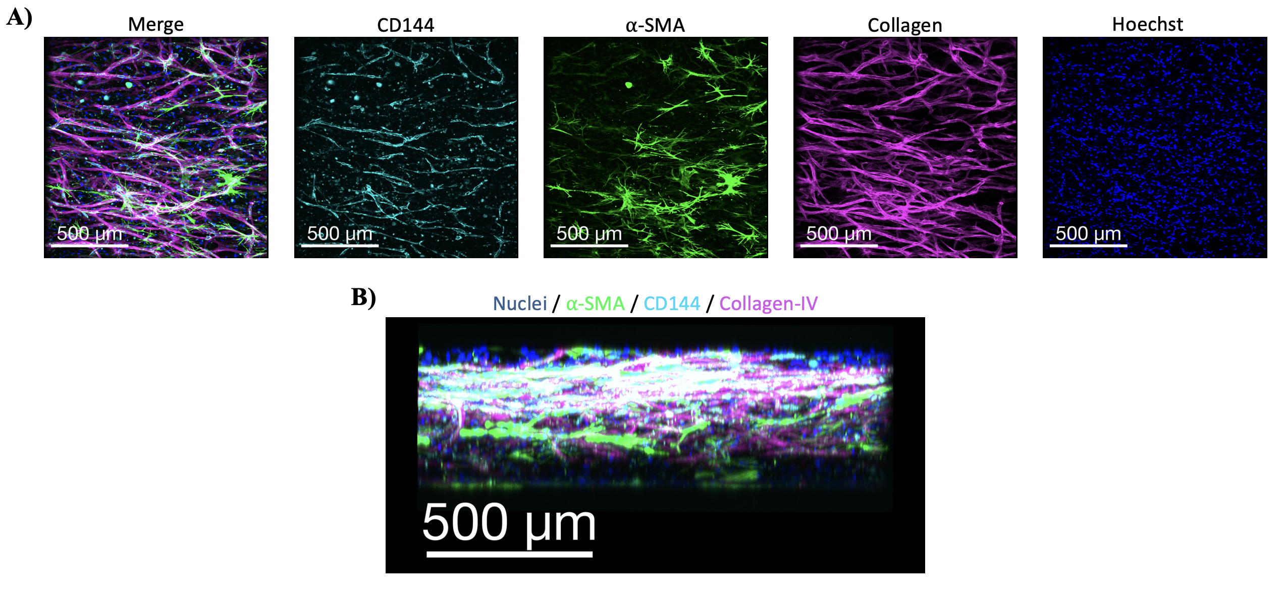
Figure 2: Co-culturing HUVECs with hMSCs in fibrin gel and 0.5 microporous membranes supports the formation of vascular structures in the basal compartment of the μSiM-oBRB chip. A) Maximum intensity projections of confocal images show the formation of distinct vascular structures by day 14. Samples were stained to visualize HUVECs (CD144; teal), 𝛼-SMA (green), Collagen-IV (pink) and nuclei (Hoechst; blue). B) Cells are well spread throughout the 3D fibrin matrix.
We set out to vascularize a μSiM-oBRB chip to model healthy and diseased maculae by co-culturing HUVECs and hMSCs in the chip’s basal channel to mimic the choriocapillaris and growing an ARPE monolayer above the porous membrane. We observed that the cells in the nanoporous membranes took longer to spread out after seeding when compared to those in the 0.5 microporous membranes and dual scale membranes. This was expected since the nanoporous membrane has significantly smaller pores and is the least permeable to media. We observed distinct vasculature spread out in the fibrin matrix that was maintained in the 0.5 microporous membranes by day 14 (Fig. 2). Immunostaining for VE-cadherin (CD144), collagen-IV, 𝛼-SMA and nuclei confirmed that the HUVECs actively deposited collagen-IV as they remodel the matrix in addition to organizing into tubular structures (Davis & Kemp, 2023). This is evidenced by the co-localization of VE-Cadherin and Collagen-IV. Additionally, the hMSCs formed supportive pericytes adjacent to developing vascular structures to provide mechanical support and prevent collapse as seen with the 𝛼-SMA expression. Collectively, these data indicate functional maturation of microvascular structures.
In developing this procedure, we have identified several common pitfalls that we recommend avoiding. In preliminary experiments, little to no tubular organization was observed at day 14 in samples prepared with 5 mg/ml of fibrinogen prepared in PBS (pH ~7.2) and 2 U/mL thrombin, with fibrin gels contracting from the periphery of the channel. Furthermore, confocal microscopy showed that the gels were separating into two distinct layers – one right under the porous membrane and the other at the bottom of the channel, with no interaction between the two (Fig. 3). The top layer was characterized by patches of monolayers of HUVECs, and very little tubular organization as observed under the light microscope by day 7. The bottom layer comprised isolated clumps of cells, indicating cell death. This observation prompted speculation about the impact of PBS solutions and the concentrations of thrombin used on fibrin polymerization. It has been previously shown that pH can drastically affect the mechanical properties of fibrin hydrogels (Jarrell et al., 2021; Takegawa et al., 2022; Wachendörfer et al., 2023). We compared how the use of PBS (pH 7.2) vs DPBS (pH 7.4) affects the gelation of fibrin hydrogels. We observed that diluting fibrinogen in PBS (pH ~7.2) formed soft, amorphous hydrogels, while mixing fibrinogen in DPBS (pH ~7.4) supported the formation of stiffer hydrogels that retained cylindrical structure. Critically, hydrogels formed using DPBS (pH 7.4) supported vasculogenesis, while the PBS (pH 7.2) fibrin gels did not (Fig. 2, Fig. 3). These observations support our hypothesis that pH affects the polymerization of fibrin hydrogels and must be controlled when forming 3D vessels in chips.
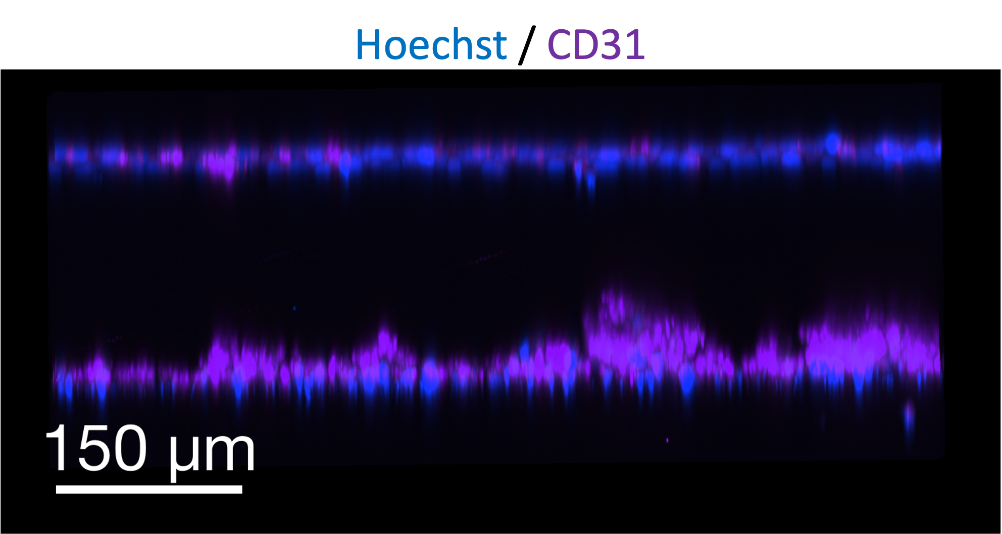
Figure 3: Uneven polymerization of fibrin results in two distinct layers. Two distinct layers are formed from the fibrin made using PBS and 100 U/ml thrombin stock by day 14. Samples were stained with Hoechst (nuclei; blue), CD31 (endothelial cell marker, purple), anti- 𝛼 SMA and 𝛼-Collagen-IV for confocal microscopy. anti-𝛼 SMA and 𝛼-Collagen-IV are not shown for simplicity.
Further, we hypothesized that uneven fibrin gel polymerization was causing gel contraction and inhibiting cell adhesion. Thrombin is inherently fast-acting; it can polymerize fibrinogen to make blood clots within 12–19 seconds (Turley et al.). Our stock solution of thrombin was very concentrated (100 U/ml), meaning we were initially mixing 2 μl of 100 U/ml thrombin into 100 μl of 5 mg/ml fibrinogen cell suspension to get a final thrombin concentration of 2 U/ml. We speculated that this highly concentrated volume of fast acting thrombin polymerized the fibrinogen molecules within its immediate surroundings, rather than react evenly with the bulk of the gel solution. We observed that fibrin hydrogels clogged the pipette tips while injecting thrombin into the final gel precursor solution before it could be fully mixed. Consequently, a fraction of the cells was incorporated into the fibrin gel, and the remaining were suspended in unpolymerized fibrinogen and later died during the maintenance period. To rectify this issue, we modified the gel formation protocol as follows. Cells were first resuspended in diluted thrombin (4 U/mL stock diluted in DPBS). Next, fibrinogen (10 mg/mL) was added to the pre-diluted mixture of cells, thrombin, and DPBS to achieve the final concentrations specified in the Methods section. Using a more diluted stock of thrombin helps moderate the rapid polymerization reaction by reducing the local concentration and immediate availability of thrombin molecules, providing enough time to thoroughly mix the samples and seed the devices. Following this adaptation, the cells distributed within the 3D fibrin matrix and developed distinct and dense vascular structures by day 14 (Fig. 2).
Altogether, we have developed a robust protocol for seeding self-assembling microvasculature in the basal channel of µSiM devices. Next, we will culture an ARPE-19 monolayer on top of the porous membrane to re-capitulate the RPE-choriocapillaris interface in the oBRB. Once we successfully produce a model of the oBRB, we will spike the apical media with VEGF induce choroidal neovascularization and migration of HUVECs into the apical well through microporous membranes. This will result in the choriocapillaris breaking through the porous membrane and ARPE-19 monolayer into the region normally occupied by photoreceptors mimicking the angiogenic environment of AMD.
In conclusion, these efforts lay the foundation for a robust and physiologically relevant oBRB model that simulates healthy and diseased retinas. In completing this work, we expect to establish a reproducible platform for studying AMD mechanisms and evaluating therapeutic interventions.
Acknowledgements
This project was completed as part of Rufaro Gamariel’s rotation project in Dr. James McGrath’s lab. The authors thank Dr. Venktesh Shirure and Dr. Steven C. George for providing the vascularization protocol and technical support that was adapted for this project.
References
- Abcouwer, S. F., Miglioranza Scavuzzi, B., Kish, P. E., Kong, D., Shanmugam, S., Le, X. A., Yao, J., Hager, H., & Zacks, D. N. (2024). The mouse retinal pigment epithelium mounts an innate immune defense response following retinal detachment. Journal of Neuroinflammation, 21(1), 74. https://doi.org/10.1186/s12974-024-03062-2
- Davis, G. E., & Kemp, S. S. (2023). Extracellular Matrix Regulation of Vascular Morphogenesis, Maturation, and Stabilization. Cold Spring Harb Perspect Med, 13(4). https://doi.org/10.1101/cshperspect.a041156
- Jarrell, D. K., Vanderslice, E. J., Lennon, M. L., Lyons, A. C., VeDepo, M. C., & Jacot, J. G. (2021). Increasing salinity of fibrinogen solvent generates stable fibrin hydrogels for cell delivery or tissue engineering. PLOS ONE, 16(5), e0239242. https://doi.org/10.1371/journal.pone.0239242
- Jarvis, G. E., Atkinson, B. T., Frampton, J., & Watson, S. P. (2003). Thrombin-induced conversion of fibrinogen to fibrin results in rapid platelet trapping which is not dependent on platelet activation or GPIb. Br J Pharmacol, 138(4), 574–583. https://doi.org/10.1038/sj.bjp.0705095
- Kurokawa, Y. K., Yin, R. T., Shang, M. R., Shirure, V. S., Moya, M. L., & George, S. C. (2017). Human Induced Pluripotent Stem Cell-Derived Endothelial Cells for Three-Dimensional Microphysiological Systems. Tissue engineering. Part C, Methods, 23(8), 474–484. https://doi.org/10.1089/ten.TEC.2017.0133
- Lejoyeux, R., Benillouche, J., Ong, J., Errera, M.-H., Rossi, E. A., Singh, S. R., Dansingani, K. K., da Silva, S., Sinha, D., Sahel, J.-A., Freund, K. B., Sadda, S. R., Lutty, G. A., & Chhablani, J. (2022). Choriocapillaris: Fundamentals and advancements. Progress in Retinal and Eye Research, 87, 100997. https://doi.org/https://doi.org/10.1016/j.preteyeres.2021.100997
- Moura, J. A., Barlow, H. J., Doak, S. H., Hawkins, K., Muller, I., & Clift, M. J. D. (2024). Exploring the Role of Fibrin Gels in Enhancing Cell Migration for Vasculature Formation. J Funct Biomater, 15(9). https://doi.org/10.3390/jfb15090265
- Nguyen, K. H., Patel, B. C., & Tadi, P. (2023). Anatomy, Head and Neck: Eye Retina. In StatPearls (Ed.), StatPearls [Internet]. StatPearls Publishing.
- Rodriguez-Cruz, J. J., Cutrufello, J., Lam, M., Nallaparaju, S., & Peppas, N. A. (2025). Drug Delivery Technologies for the Treatment of Age-Related Macular Degeneration. Adv Sci (Weinh), 12(39), e03212. https://doi.org/10.1002/advs.202503212
- Shirure, V. S., Hughes, C. C. W., & George, S. C. (2021). Engineering Vascularized Organoid-on-a-Chip Models. Annual Review of Biomedical Engineering, 23(1), 141–167. https://doi.org/10.1146/annurev-bioeng-090120-094330
- Shirure, V. S., Lezia, A., Tao, A., Alonzo, L. F., & George, S. C. (2017). Low levels of physiological interstitial flow eliminate morphogen gradients and guide angiogenesis. Angiogenesis, 20(4), 493–504. https://doi.org/10.1007/s10456-017-9559-4
- Song, M. J., Quinn, R., Nguyen, E., Hampton, C., Sharma, R., Park, T. S., Koster, C., Voss, T., Tristan, C., Weber, C., Singh, A., Dejene, R., Bose, D., Chen, Y.-C., Derr, P., Derr, K., Michael, S., Barone, F., Chen, G.,…Bharti, K. (2023). Bioprinted 3D outer retina barrier uncovers RPE-dependent choroidal phenotype in advanced macular degeneration. Nature Methods, 20(1), 149–161. https://doi.org/10.1038/s41592-022-01701-1
- Summer, S., Rossmanith, E., Pasztorek, M., Fiedler, C., Gröger, M., Rauscher, S., Weber, V., & Fischer, M. B. (2022). Mesenchymal stem cells support human vascular endothelial cells to form vascular sprouts in human platelet lysate-based matrices. PLoS One, 17(12), e0278895. https://doi.org/10.1371/journal.pone.0278895
- Takegawa, Y., Takao, T., Sakaguchi, H., Nakai, T., Takeo, K., Morita, Y., Toyonaga, T., & Kodama, Y. (2022). The importance of pH adjustment for preventing fibrin glue dissolution in the stomach: an in vitro study. Scientific Reports, 12(1), 6986. https://doi.org/10.1038/s41598-022-10968-5
- Turley, R. K., Sather, R., & Zercher, R. Thrombin Time. Retrieved 1 November fromhttps://www.urmc.rochester.edu/encyclopedia/content?contenttypeid=167&contentid=thrombin_time
- van Duinen, V., Stam, W., Mulder, E., Famili, F., Reijerkerk, A., Vulto, P., Hankemeier, T., & van Zonneveld, A. J. (2020). Robust and Scalable Angiogenesis Assay of Perfused 3D Human iPSC-Derived Endothelium for Anti-Angiogenic Drug Screening. International Journal of Molecular Sciences, 21(13), 4804.
- Wachendörfer, M., Buhl, E. M., Messaoud, G. B., Richtering, W., & Fischer, H. (2023). pH and Thrombin Concentration Are Decisive in Synthesizing Stiff, Stable, and Open-Porous Fibrin-Collagen Hydrogel Blends without Chemical Cross-Linker. Adv Healthc Mater, 12(10), e2203302. https://doi.org/10.1002/adhm.202203302
Well done Rufaro and Kevin. High standard on this post. Looking forward to Part II.