Theoretical Underpinnings of Small Molecule Permeability Measurements in the µSiM (Part 3: Application to Cell Barriers)*
This is the third installment in a series of posts focused on the development of a method for measuring barrier permeability in the µSiM. In Part 1 we discussed the goal of making these measurements in situ at a position beneath the membrane. In Part 2 we validated this approach experimentally on the Andor Dragonfly Spinning Disk microscope. As part of this validation we were able to accurately measure the free diffusion of Alexa488 dextran with excellent quality-of-fit to the solution to the free diffusion equation when we made measurements at 100 µm below the membrane. In this post will see that the application to cell permeability requires a different equation, but that if we get the physics of the situation right, the in situ method again works.
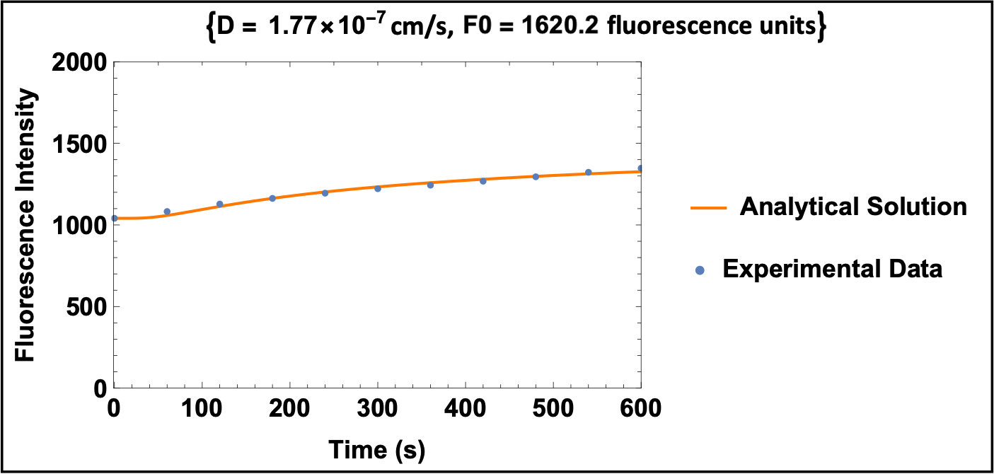
First a look at the bad fits to cellular data and the free diffusion equation…
When we try our two parameter method, while the analytical solution fits the data well, the source fluorescence intensity (F0), is far lower than measured (1620.2 fit compared to 6735 measured fluorescence units). This tells us the analytical solution is not a good representation of the experimental data.
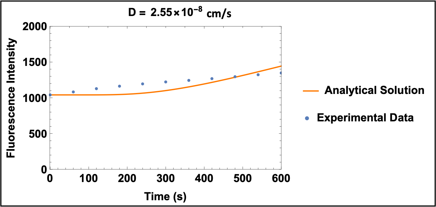
However, when we switch to our one parameter fit, inputting F0 and only solving for the diffusion coefficient, we get a really poor fit to the data.
Our original idea here was to find a new ‘hindered’ diffusion coefficient and then figure out how to interpret it as a cellular permeability. The fact that the fits were terrible was the math’s way of saying our idea was terrible too. An alternative and more physiological way to view the situation is to assume that cells are controlling the rate of molecular transport across them by maintaining a constant flux. This assumption is implicit to all diffusion-based measurements of permeability in the literature. In this case the boundary condition we need to use in solving the diffusion equation is not constant concentration at the membrane , but a constant flux at the membrane which we will call Jo. This picture looks like this …
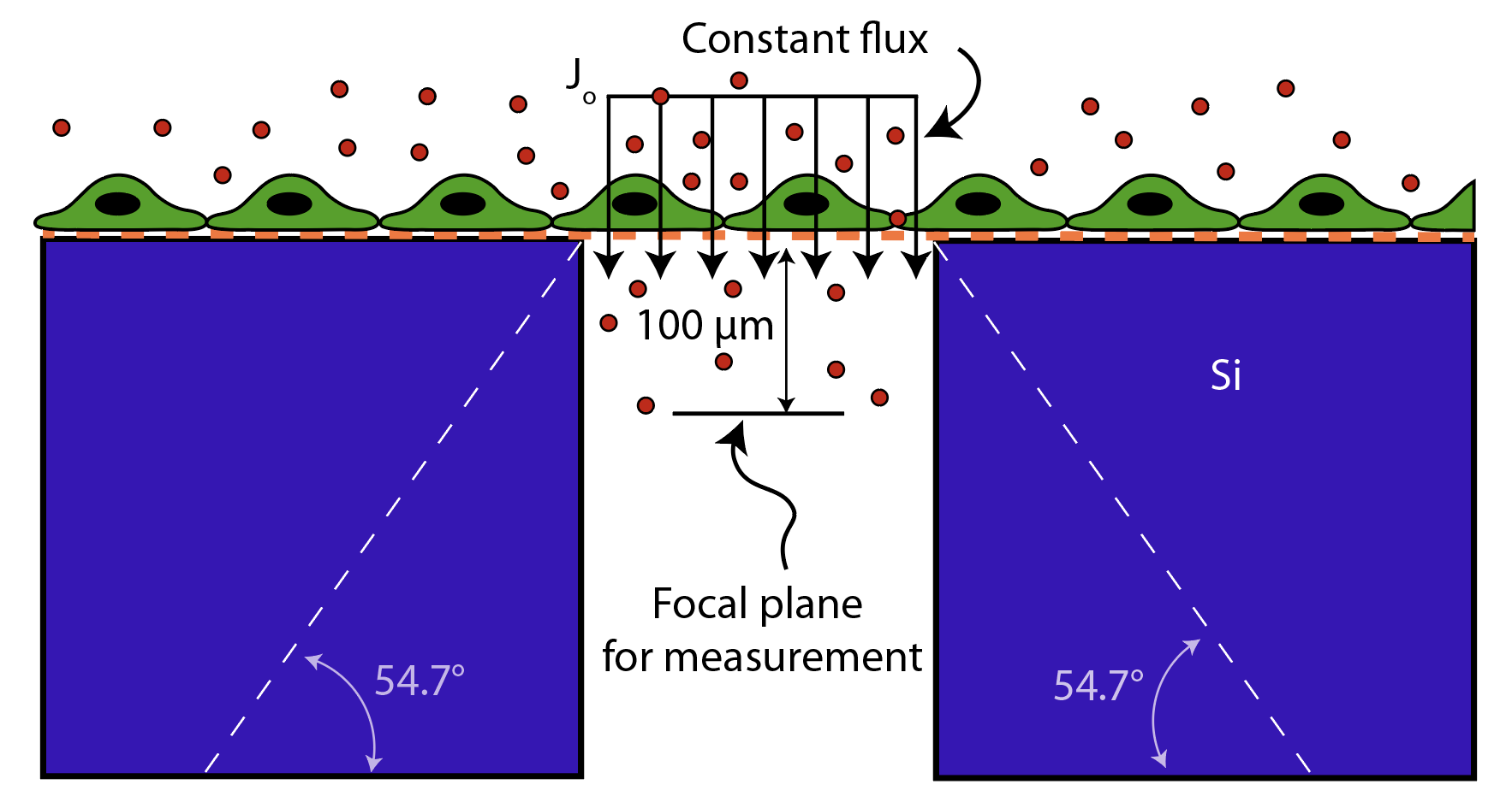
*For the purposes of our work here (and in the case of free diffusion in Part 2) we will ignore the expansion of the opening in the trench which is illustrated with the dashed lines (we will come back and estimate the error associated with that expansion later).
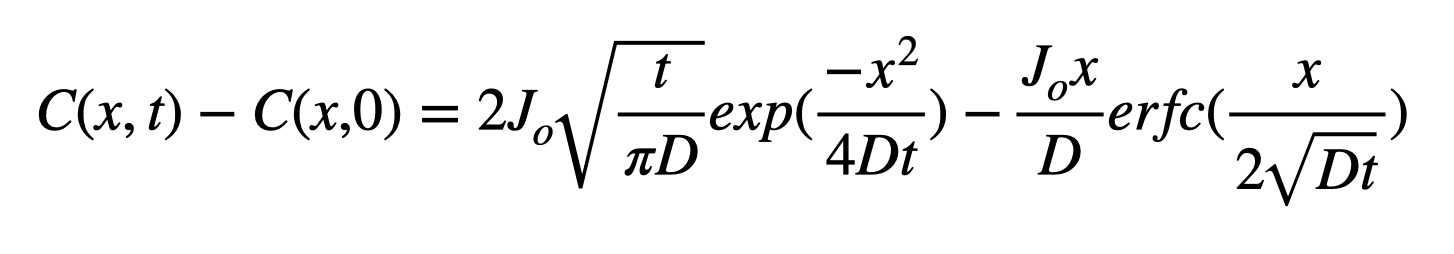
The classic solution to 1D diffusion into a semi-infinite space with a constant flux boundary condition at x = 0 is given by …
 And the permeabilty is related to the flux by …
![]()
So if we normalize the transport equation by a concentration, the flux term becomes the permeability.
We will convert concentrations to fluorescence in the usual manner and use the same normalization we did for free diffusion in Part 2 to get …
 This is the equation to be fit to our data. Note that D is the free diffusion coefficient which we will need to either grab from literature, calculate based on a Stokes-Einstein approximation, and/or measure ourselves as in Part 2.
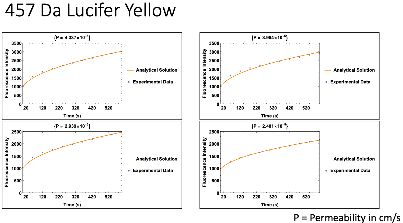
This approach was validated for our three fluorescent dyes (10 kDa FITC-Dextran, 10 kDa Dextran conjugated to AF488, and 457 Da Lucifer Yellow), comparing results across on hCMEC/D3 monolayer to permeabilities reported in the literature. The equation above was used, with Pe as the only free parameter. The diffusion coefficient for each molecule was obtained from literature.
Here are example fits for each dye:
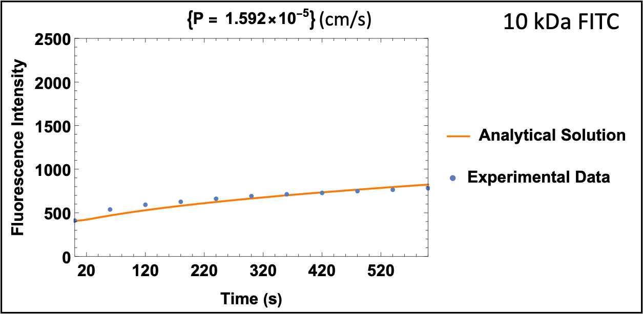

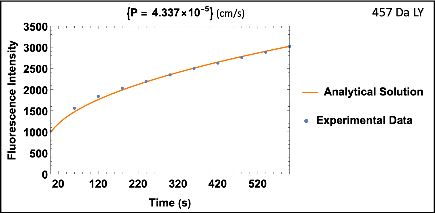
The permeability values for well established dyes match well to values found the in literature for 10 kDa Dextran, however, Lucifer Yellow data may be slightly off:
| hCMEC Permeability | 10 kDa FITC Dextran | 10 kDa Dextran-AF488 | 457 Da Lucifer Yellow |
| Experimental |
1.33 ± 0.26 x 10^-5 cm/s 0.80 ± 0.15 x 10^-3 cm/min |
3.13 ± 0.71 x 10^-6 cm/s 1.88 ± 0.43 x 10^-4 cm/min |
3.42 ± 0.90 x 10^-5 cm/s 2.05 ± 0.54 x 10^-3 cm/min |
| Literature |
0.5 – 3.4 x 10^-5 cm/s 0.2 – 2.0 x 10^-3 cm/min |
Unknown |
1.0 – 2.6 x 10^-5 cm/s 0.6 – 1.6 x 10^-3 cm/min |
10 kDa FITC Dextran: References 1-2; 457 Da Lucifer Yellow: References 3-5. All references are +hydrocortisone/-LiCl. Some are apparent permeability, whereas others are endothelial permeability.
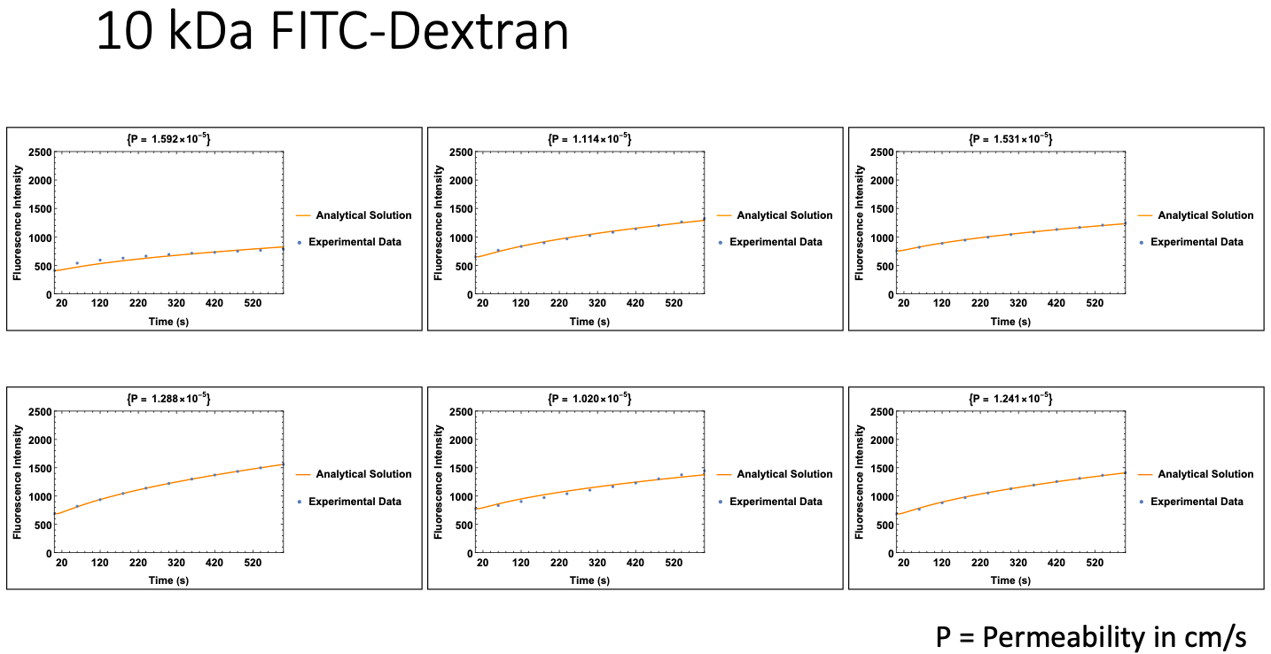
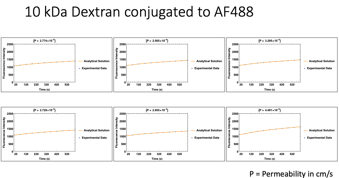
Here are the fits for the rest of the data, showing some (but minimal) variability:
For those interested, this is the code used in Mathematica:
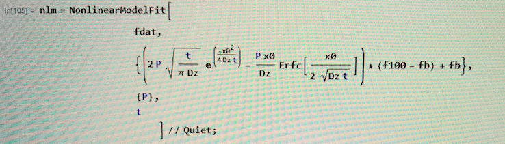
This is just rearranged from the code shown above, so the raw data (Fx,t or fdat) can be inputed and permeability automatically calculated. Dz is the diffusion coefficient of the fluorescent dye, x is 0.1 mm (distance below membrane, UPDATE: we now use 0.133 mm due to the “fishtank effect“), f100 is the fluorescence intensity of the source concentration, and fb is background fluorescence.
NOTE: THIS POST CURRENTLY SUFFERS FROM A PROLIFERATION OF NAMES FOR THE SAME THINGS.
- The final fluorescence expected is called F_inf in the theory but F0 in the figures and F100 in the code.
- The permeability is called P in the figures and code but Pe in the theory.
- Thankfully, the background flourescence is called Fb everywhere.
Bibliography
- Yang, Z. et al. Autophagy Protects the Blood-Brain Barrier Through Regulating the Dynamic of Claudin-5 in Short-Term Starvation. Frontiers in Physiology 10, 2 (2019).
- Förster C, Burek M, Romero IA, Weksler B, Couraud PO, Drenckhahn D. Differential effects of hydrocortisone and TNFalpha on tight junction proteins in an in vitro model of the human blood-brain barrier. J Physiol. 2008;586(7):1937-1949. doi:10.1113/jphysiol.2007.146852
- Weksler B, Romero IA, Couraud PO. The hCMEC/D3 cell line as a model of the human blood brain barrier. Fluids Barriers CNS. 2013 Mar 26;10(1):16. doi: 10.1186/2045-8118-10-16. PMID: 23531482; PMCID: PMC3623852.
- Eigenmann, D.E., Xue, G., Kim, K.S. et al. Comparative study of four immortalized human brain capillary endothelial cell lines, hCMEC/D3, hBMEC, TY10, and BB19, and optimization of culture conditions, for an in vitro blood–brain barrier model for drug permeability studies. Fluids Barriers CNS 10, 33 (2013). https://doi.org/10.1186/2045-8118-10-33
- Bhupathiraju, N. V., Hu, X., Zhou, Z., Fronczek, F. R., Couraud, P. O., Romero, I. A., Weksler, B., & Vicente, M. G. (2014). Synthesis and in vitro evaluation of BBB permeability, tumor cell uptake, and cytotoxicity of a series of carboranylporphyrin conjugates. Journal of medicinal chemistry, 57(15), 6718–6728. https://doi.org/10.1021/jm500786c