
Human brain vascular pericyte viability in endothelial media
Introduction
Mural cells, including smooth muscle cells and pericytes, are important for supporting endothelial cells in the vasculature [1]. They play a role in angiogenesis, and contribute to the development of the vascular basement membrane that provides structure for blood vessels [1,2]. Numerous in vitro models of the blood-brain barrier (BBB) have incorporated pericytes alongside endothelial cells to closely mimic their physiological arrangement [2-4]. Previously, we arranged human cerebral microvascular endothelial cells (hCMEC/D3) and primary human brain vascular pericytes (HBVP) on either side of the μSiM-hNVU’s nanoporous membrane to model the BBB. In these earlier experiments, HBVPs were first seeded in pericyte media and were maintained in this fashion for 1 or 2 days prior to hCMEC/D3 seeding. Once the endothelial cells were seeded, media in the μSiM devices was switched over to the hCMEC assay media. While it appeared that HBVPs survived throughout the experimental period, it was unclear how much the media switch affected their viability.
Method
HBVPs (ScienCell, cat. no. 1200) at passage 7 were seeded in a 24-well plate in pericyte media +2% FBS + 1% pericyte growth supplement + 1% penicillin/streptomycin (ScienCell, cat. no. 1201) at a density of ~5000 cells/cm2. The plate had previously been coated with 2 μg/cm2 poly-L-lysine (ScienCell cat. no. 0403) to promote HBVP adhesion, and was kept at 4°C until seeding. This plate was incubated at 37°C and 5% CO2. After 2 days, the media was exchanged for either pericyte media, hCMEC assay media (EBM-2 +0.4% hFGF +0.04% hydrocortisone +0.1% Gentamycin +2% FBS; Lonza, cat. no. CC-3162), or a 50/50 mix of the two media. PBS was utilized as a control. The cells were maintained for 7 days, with media exchanges performed every 2-3 days. A live/dead stain was then performed using calcein-AM and ethidium homodimer-1 mixed in pericyte media. Following 15 min of incubation with the stains, the cells were imaged using a Keyence BZ-X microscope. The images were then utilized to perform a rough estimate of viability expressed as the percentage of calcein-AM-stained nuclei out of the total number of stained nuclei (following the tutorial available here).
Results
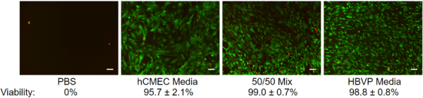
Viability was highest for HBVPs cultured in pericyte media or the 50/50 media mix (~99%). Cells cultured in hCMEC assay media had a slightly lower viability (~96%) and appeared to be more spread than cells cultured in the other media conditions. No cells survived culture in PBS.

Discussion
Even though the HBVPs appeared to have lower viability in hCMEC assay media compared to the other conditions, they still tended to have very high viability (in excess of 90%). We are not concerned about the effects of the media exchange in terms of HBVP survivability. However, it still remains to be seen whether culture in the endothelial media affects the expression of characteristic pericyte markers such as NG2 and PDGFRβ [5,6]. Furthermore, it is unknown why the cells appeared to take on different morphologies in the different media mixtures. Spreading would presumably help increase coverage of endothelial tubes by HBVPs, so this behavior may actually be beneficial in a vascular model [2].
References
[1] K. Gaengel, G. Genové, A. Armulik, and C. Betsholtz, “Endothelial-Mural Cell Signaling in Vascular Development and Angiogenesis,” Arteriosclerosis, Thrombosis, and Vascular Biology, vol. 29, pp. 630-638, 2009/05/01 2009.
[2] A. N. Stratman, K. M. Malotte, R. D. Mahan, M. J. Davis, and G. E. Davis, “Pericyte recruitment during vasculogenic tube assembly stimulates endothelial basement membrane matrix formation,” Blood, vol. 114, pp. 5091-5101, 2009.
[3] M. S. Thomsen, S. Birkelund, A. Burkhart, A. Stensballe, and T. Moos, “Synthesis and deposition of basement membrane proteins by primary brain capillary endothelial cells in a murine model of the blood–brain barrier,” Journal of Neurochemistry, vol. 140, pp. 741-754, 2017/03/01 2017.
[4] Y. Yamazaki, et al., “ApoE (Apolipoprotein E) in Brain Pericytes Regulates Endothelial Function in an Isoform-Dependent Manner by Modulating Basement Membrane Components,” Arteriosclerosis, Thrombosis, and Vascular Biology, vol. 40, pp. 128-144, 2020/01/01 2020.
[5] J. Fukushi, I. T. Makagiansar, and W. B. Stallcup, “NG2 Proteoglycan Promotes Endothelial Cell Motility and Angiogenesis via Engagement of Galectin-3 and α3β1 Integrin,” Molecular Biology of the Cell, vol. 15, pp. 3580-3590, 2004/08/01 2004.
[6] K. K. Hirschi, S. A. Rohovsky, and P. A. D’Amore, “PDGF, TGF-β, and Heterotypic Cell–Cell Interactions Mediate Endothelial Cell–induced Recruitment of 10T1/2 Cells and Their Differentiation to a Smooth Muscle Fate,” Journal of Cell Biology, vol. 141, pp. 805-814, 1998.
Update: NG2 and PDGFRb Staining
