Ongoing Human Pulmonary Microvascular Endothelial Cell Culture and Characterization
Introduction
In an effort to move forward with a ‘lung microvasculature on a chip’ model, I have been experimenting with using primary human pulmonary microvascular endothelial cells in our devices. The cells (HPMECs) are purchased from Promocell and are collected from a single donor lung. Unfortunately I found little information on their use in recent literature, so this preliminary work is focused on characterizing cell phenotype and health in culture.
Results 1 – Coatings and Device Configuration
Of note, in standard flask culture, these cells double much slower than HUVECs (~35 h vs 12 h). Moving to closed-top flow devices and starting with what we know best, 100 nm NPN membranes were coated with .17 mg/ml human fibronectin and HPMECs were perfused. After 24 h culture, few cells remained adhered, with all cells dying at 48 h (Figure 1).

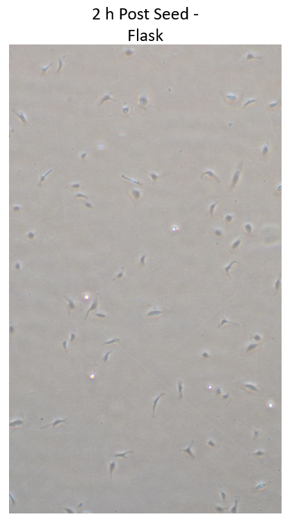
Following a suggestion from the Waugh lab, 100 nm NPN membranes were coated with 5 ug/cm^2 collagen I, and cells were perfused. Initially, HPMECs adhered very well to the altered coating, however, cells started dying at 24 h, and were completely gone at 48 h (Figure 2, 48 h not shown). Cells at 2 h in a standard flask showed similar phenotype to those in the device, so this strongly suggests something in the device/associated with the device is killing the cells over the 24 h post seed (Figure 3, cells in flask grow fine*).

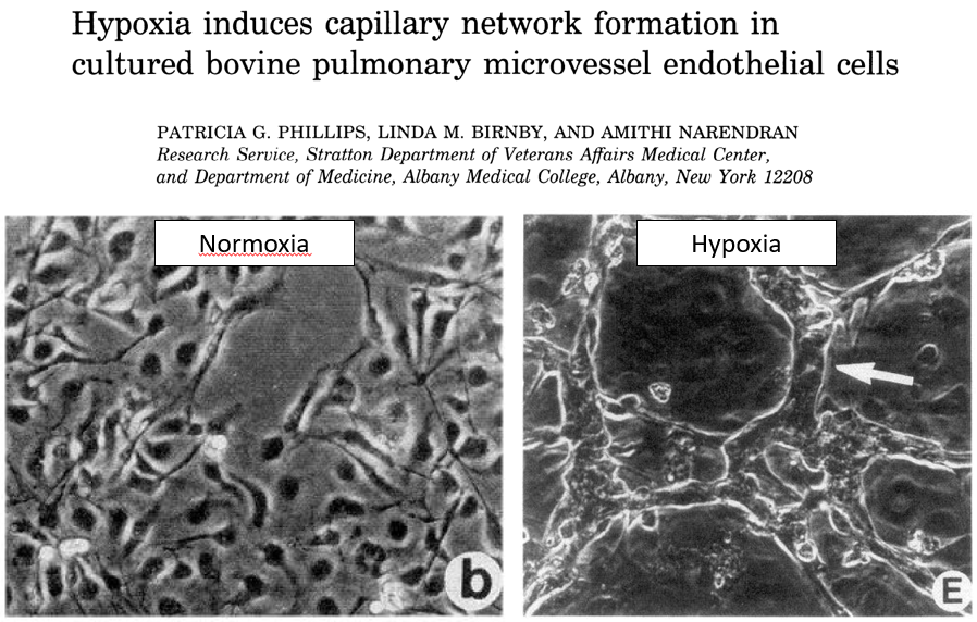
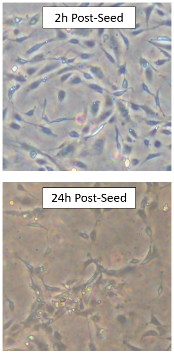
After further discussion with Jim, we talked about moving to an open-top device, as gas exchange may be an issue in our close-top flow devices, potentially causing the cell death in extended culture. After some more digging, I was able to find a paper that explored the effects of hypoxia (decreased oxygen) on bovine pulmonary microvascular endothelial cells (Figure 4). Interestingly, when compared to my experiments with the successful coatings, the cell phenotype looks much like the hypoxic cells in the aforementioned paper (Figure 5).
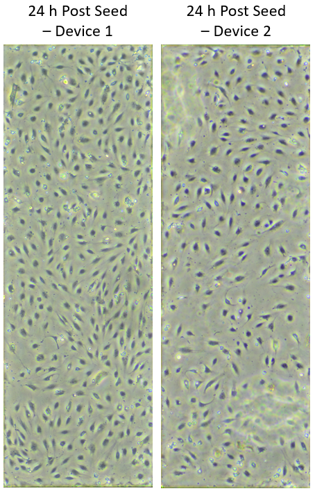
These results support the pour gas exchange hypothesis, so we would expect open-top devices to fix these issues. Again, 100 nm NPN membranes were coated with 5 ug/cm^2 collagen 1, and HPMECs were seeded. As expected, cells are looking much healthier 24 h post-seeding (Figure 6).
Conclusion 1
Collagen I coatings support healthy HPMEC adherence, and open-top devices allow for sufficient gas exchange. Images of open and closed top devices are below for those not familiar (Figure 7).

Results 2 – Characterization and PMN Migration
Now that we can achieve stable HPMEC culture in uSiMs, I wanted to characterize EC markers, as well as try some preliminary neutrophil (PMN) migration assays to see the results compare to HUVECs. The main concern isn’t necessarily the cell change, but instead the change of device format.
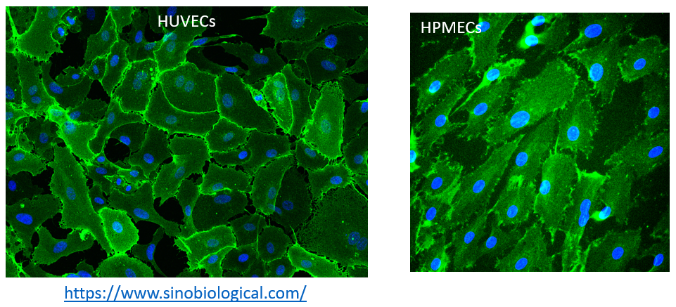
Initially, I stained confluent HPMECs for CD31 (PECAM-1) to confirm EC lineage (Figure 8).

After confirmation of cell lineage, I ran a quick PMN migration experiment with modification for open-top devices. PMNs were isolated as usual, but cells were add in bolus to the top well of the uSiM and incubated for 15 mins at 37C and 5% CO2. Next, 10 nM fMLP was perfused into the bottom channel and images were collect (phase, 8 fpm).

Conclusion 2
HPMECs stain positive for EC marker CD31. PMNs migration through following fMLP as expected.