Immunogold Labeling of Exosomes
So, this is the data that we have all been waiting for. Without all the fancy introductions and stuff, I present you with labeled exosomes. They were isolated in tangential flow from my human plasma samples using the SOP that I have been following along with a labeling protocol that Henry and I developed. It is as follows:
- Capture the exosomes on the membrane. Remove it from the device.
- Wash with 3 drops of PBS (drops are defined as being 50 μL in volume in every step).
- Bind anti-CD63 antibody (2 μg/mL) for 40 min (incubate on 50 μL drop).
- Wash with 3 drops of 0.1 wt% casein in PBS.
- Bind streptavidin immunogold secondary (1:200 concentration) for 40 min (incubate on 50 μL drop).
- Wash with 3 drops of PBS.
- Wash with 5 drops of NP water.
- Remove excess water and allow to air dry.
- Coat with 3 nm of gold and image in SEM.
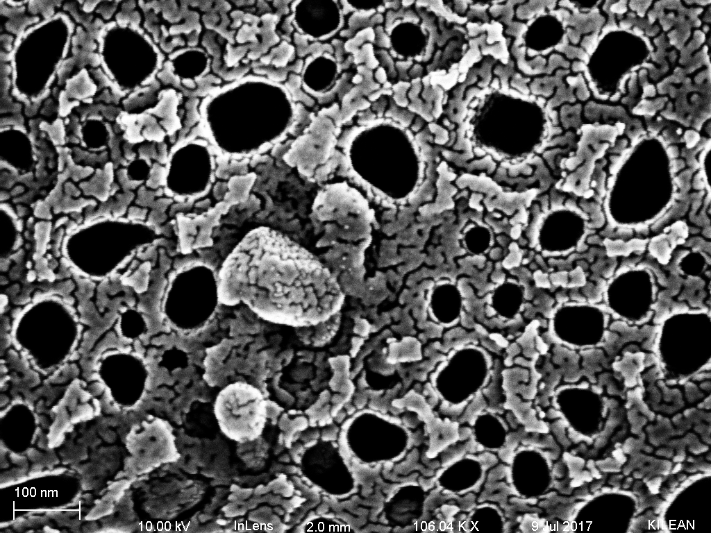
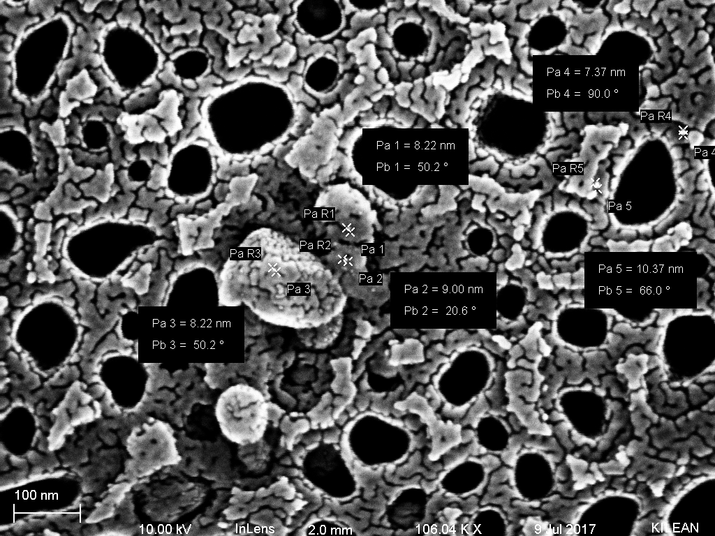
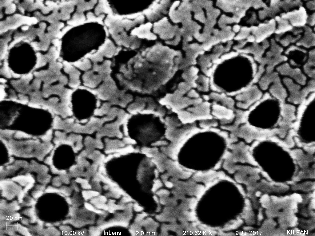
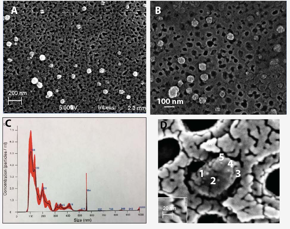
As we can see from the images below, there are particles bound to the surface of the exosomes that are the correct size for the gold nanoparticles in the immunogold label.

Now that I have these images, as well as a lyophilized exosome standard sample, I will be repeating the experiment to further confirm (statistics, right?) that these are indeed exosomes, but for now I think that I can safely say these are labeled exosomes and what we are capturing are indeed exosomes.