I’ve been gathering data for a paper using my microporous MgF2 nanomembranes so here are some sample figures for such a paper.
Herein, we are trying to create a porous, Raman-compatible substrate for use in cell culture. Researchers have used Raman microspectroscopy to perform quantitative non-invasive measurements of biomolecules in cells. It’s a very exciting technique. While silicon based substrates have excellent properties for cell culture, they have strong Raman background signals and absorb a lot of laser power at the illumination wavelengths we would like to use. MgF2, Quartz, and CaF2 are considered Raman-compatible for their substantially weaker backgrounds. Researchers currently deposit or culture cells on non-permeable coverglasses made of these materials. However, we have known that cells sometimes require a permeable substrate to adopt morphologies that are more consistent with in vivo studies of morphology. So using our microporous nitride substrates, we wish to create a substrate that will have both the permeability and the Raman-compatibility necessary to facilitate more accurate Raman studies of those types of cells.
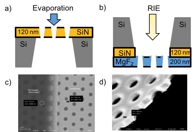
Figure 1. MgF2 nanomembrane material properties. (a)-(b) MgF2 relief pattern transfer, with cartoon crossections of microporous films (not to scale). Beginning with a freestanding film of microporous silicon nitride (a), MgF2 is evaporated onto the substrate (200 nm, 0.1-0.3 nm/sec, 250 °C, Platen Rotation), coating the porous substrate, resulting in a hybrid material. (b) The substrate is then inverted and purified using RIE (90% CHF3, 10% Oxygen, 75 mTorr, 100 W), releasing a freestanding nanoporous film of MgF2. (c) A SEM image normal to the membrane plane shows the infilling effect of the direct evaporation process, as the template pores narrow from their designed 500 nm diameters to approximately 325 nm. (d) SEM crossection of a partially etched MgF2 nanomembrane (with some template backing remaining), showing film thickness close to the targeted 200 nm.
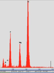
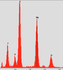
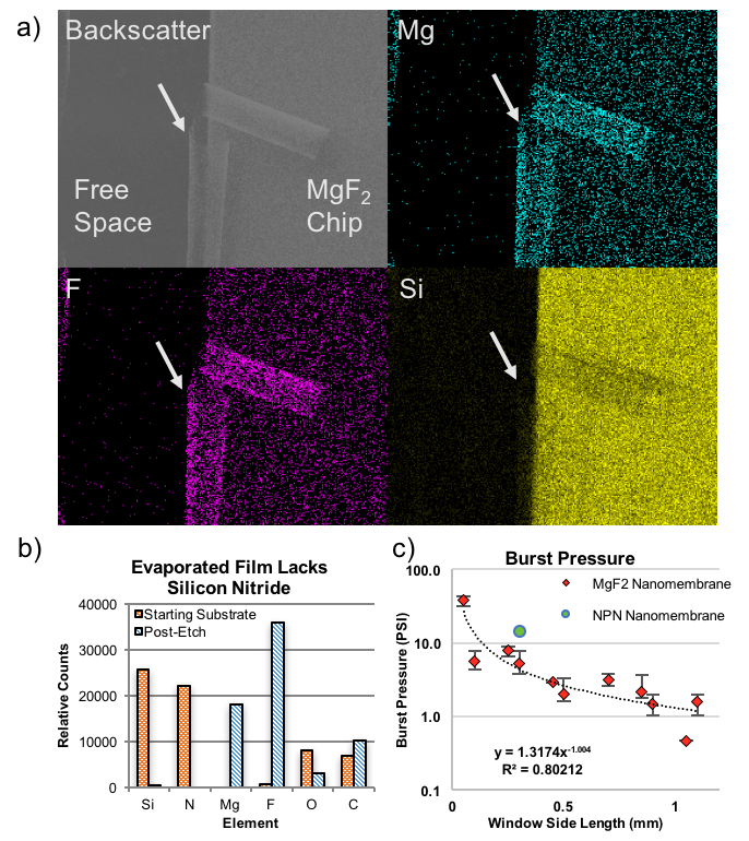
EDS measurement of the composition of the freestanding membrane. While the Mg and F signals are preserved across the freestanding region, the Si signal is not. The membranes are >85% MgF2 in the freestanding region. As a side note, there was some sample drift, so the image is blurred. This is a 30kX image.
Before Etch
After Etch
Previous Figure with 50 nm thick nanoporous MgF2. Here the burst pressure for the 200 nm microporous MgF2 is about 7 PSI, over a 5-slot structure instead of a square window.
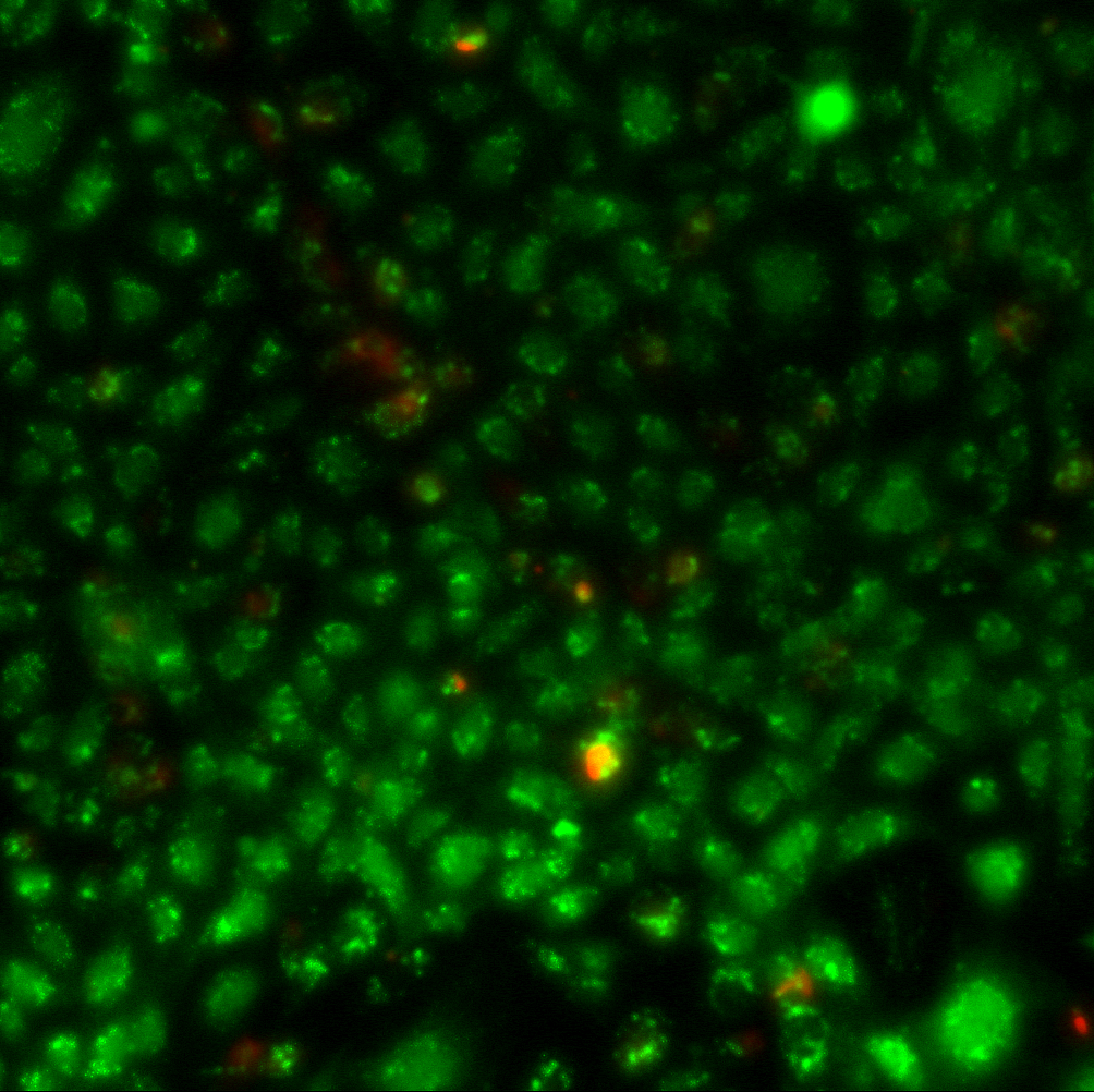
20X Live/Dead stain image of P6 HUVECS (Day 7) on fabricated microporous MgF2 grown in MCDB131 media. Eventually, we will use ARPE-19 Cells with tight junction staining which have a characteristic response to permeable substrates.
We will have to reestabilish the Raman background of the membranes, but the material composition looks to be the same as with my nanoporous material. HUVECS seem to stick ok without using any additional coatings.
In early May, the Barcikowski lab shipped us four samples of gold nanoparticles for us to run separations on. They took forever to get here, and had aggregated and precipitated by the time they arrived. About a week ago, the lab sent another batch of four samples (which differed slightly from the samples they sent…
Since the new year there have been difficulties with charging the glass plate and the charge not being enough to capture the membrane. Synthetic silk was tested to see if the glass charged better but no difference was seen between the silk and a crew wipe. When materials were first being tested to use during…
As previously stated in my initial post (found here https://trace-bmps.org/nanomembranes-in-potassium-ion-selective-electrodes), I am in the process of developing a potassium Ion Selective Electrode (ISE) Sensor using our chips. Along the way I have gathered all sorts of curious but interesting data, with observations bringing both old discoveries and new oddities to light. Outlined below are some…
I’ve run a few comparative studies with the CytoVu devices that have SiO2 membranes in them. From a previous NRG discussion, here is the comparison of a single location with only the fluorescent images, and that same location but with phase included: In the phase image, the pores can be seen through the cells, especially evident in…
It seems that pores are only formed in the “sandwich structure” on certain process. So in order to verify this conclusion, I annealed two layers structure (20nm sio2+15nm si) to test the agglomeration theory. The other parameters in depositing and annealing are same to typical sandwich structure process (1000°C anneal, ramp rate 100c/s, w/o bias,…
Cancer cell metastasis is responsible for 80 percent of cancer-related death. The metastatic cells migrate through tight constricting spaces, therefore constricted migration and its enabling mechanisms have received a lot of attentions in recent years. The most famous research groups that are working in this field are Lammerding group, Discher group, Wolf and Friedl group,…