ADSC Spreading on Various Surfaces
Overview:
The study of ADSC spreading on various surfaces was conducted to determine if porous surfaces and the porosity of the surfaces had any effect on cell spreading. Eight different surfaces were studied and each of the membranes were SiO2, these included: 0.5um low porosity (LP), 0.5um high porosity (HP), 3um LP, 3um HP, 5um LP, and non-porous, tissue culture plastic (TCP) with and without 1% Geltrex.
Methods:
ADSCs were seeded onto each membrane type as well as TCP at 300 cells/well, each surface had a sample size of 3. At 24 hours, each of the wells were fixed, permeablized and stained for Actin/Nuclei. ImageJ processes were used to make the fluorescence images binary and to then find the area of each individual cell. The area of each cell was found for each membrane type. The average cell areas used in the bar graph were found for each of the surfaces using the top 75% of the data. The data in the boxplot also only included the top 75% of data. The bottom 25% of the data was excluded to remove any dead or non-adhered cells that may have been counted.
Data:
The data was placed into a bar graph to show the comparison of average cell area. The data was also placed into a box plot to show that the data is very spread and includes many outliers.
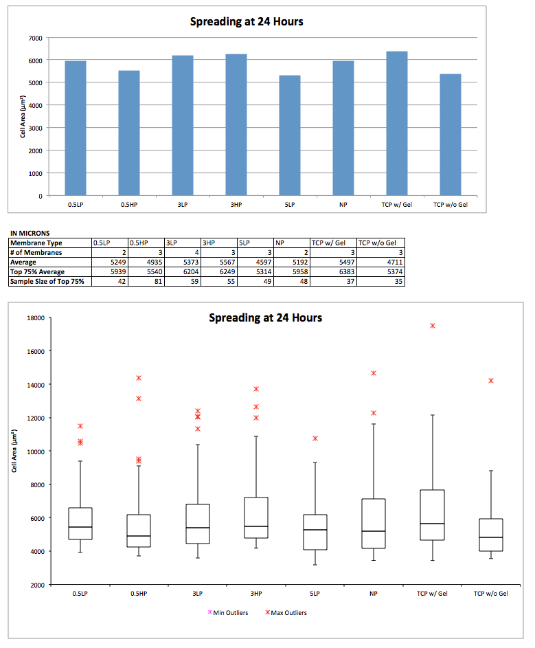
Conclusion:
After using statistical analysis, only the TCP with Geltrex and the TCP without Geltrex were statistically significantly different. The bar graph places an emphasis on each of the average ADSC areas which shows that they have very little difference. The box plot shows that for each individual surface type there is a large amount of spread and almost all of the surfaces have outliers. This shows how variable ADSC spreading can be and again supports that the surface does not effect ADSC spreading.