More Barcikowski bioconjugates have arrived. Here’s the plan for them.
The Barcikowski lab sent us generous amounts of three different samples – gold, platinum, and bioconjugated gold. Each of the samples was created via laser ablation, resulting in fairly polydisperse (4-50nm diameter) nanoparticles for the pure metal and significantly less polydisperse (2-20nm, not including bound ligand) nanoparticles in the case of bioconjugated gold. As before, the desired result is pure metal samples with a size cutoff less than 7nm (for gold) or 5nm (for platinum), and a bioconjugated sample free of unbound ligand. As before, the samples took two weeks to get here.
The Barcikowski lab included Malvern DLS scans of each of the samples. What follows are their scans of the three samples and what I was able to measure on our Malvern. Note that these are intensity scans, not the number scans I typically show.
First – our Bioconjugated Gold. Peaks are at 86.4, 2080, and 14.9nm
Theirs – peak at 118.2nm

Next – our gold. Peaks at 294, 4560, and 27.9nm 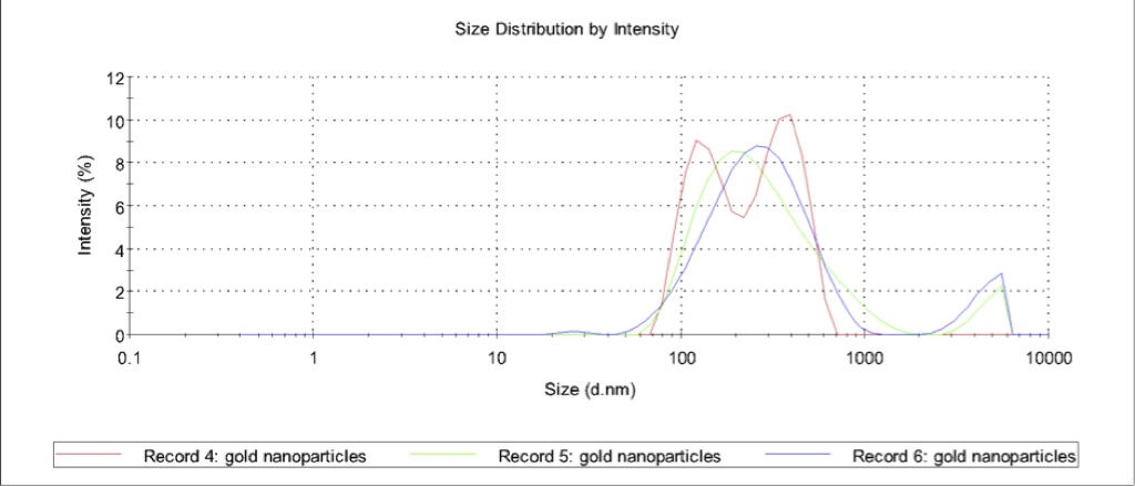
Theirs – peaks at 1417 and 177.3nm
Finally – our platinum. peak at 151nm 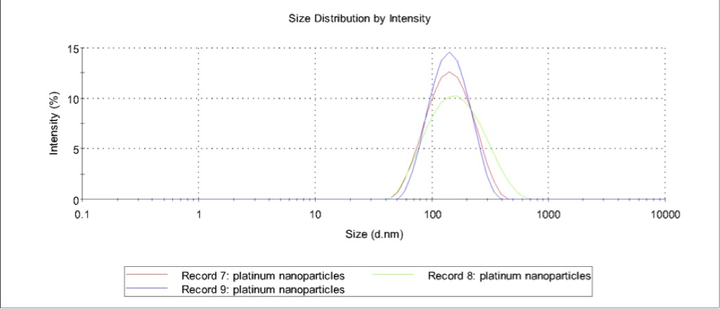
Theirs – peak s at 1803, 84.6, and 724.4nm

So pretty close, in spite of their long hot transport through customs. The only really odd thing is the Zetapotentials (graphs that the Barcikowski lab mailed us will be up in a few hours) – they measured a zetapotential of -56.4mV, +31.2mV, and +37.2, for the bioconjugated gold, gold and platinum, respectively, which contradicts both what they told us via email and the features of penetratin (see below).
The plan of attack for the Bioconjugated gold is as follows:
- Separate the gold from the free ligand in the bioconjugated sample using the Ultracentrifuge.
- Measure the relative concentration of unbound ligand in the supernate as a baseline using either simple absorbance at 280nm or a Bradford assay with a coomassie stain and a TECAN scan.
- Determine if adsorbtion to the Sepcon walls is a significant problem for the ligands by comparing the Bradford assay in (2) to that of another aliquot of spun ligand that has been pipetted into the top chamber of a membrane-less Sepcon and removed from the bottom chamber.
- Determine if adsorbtion to the Sepcon walls is a significant problem for the nanoparticles by comparing TECAN scans of two samples of 20nm gold colloids where one is flowed through a membrane-less Sepcon, as in (3).
- Perform a separation using 30nm Sepcons (Gen 3 – at least until our pressure setup is finished) on the unspun bioconjugated gold. About half of a sample of 400uL should be passed through the membrane.
- Measure the relative concentration of unbound ligand in the filtrate using the Bradford assay. With luck, the concentration of the ligand in the filtrate should be about equal to the ligand concentration in the spun sample. A TECAN measurement of the retentate should also be taken so that we can be sure that the nanoparticles aren’t making it through the membrane.
- Add enough buffer to the retentate to restore its volume to 400uL and repeat steps 4 and 5 at least three times.
For the pure metal samples, we’ll use Josh’s new large-pore PEG-lyated membranes to run a single separation of each sample and characterize the size and concentration of the retentate and supernate using both the Malvern and the TECAN.
The lab has been a little reticent about the nature of the peptide they’ve conjugated, but they recently released an article about Penetratin-conjugated gold nanoparticles, and penetratin seems to fit the scant description of the mystery peptide I’ve been given:

Their published data does match pretty closely what we’ve been seeing:
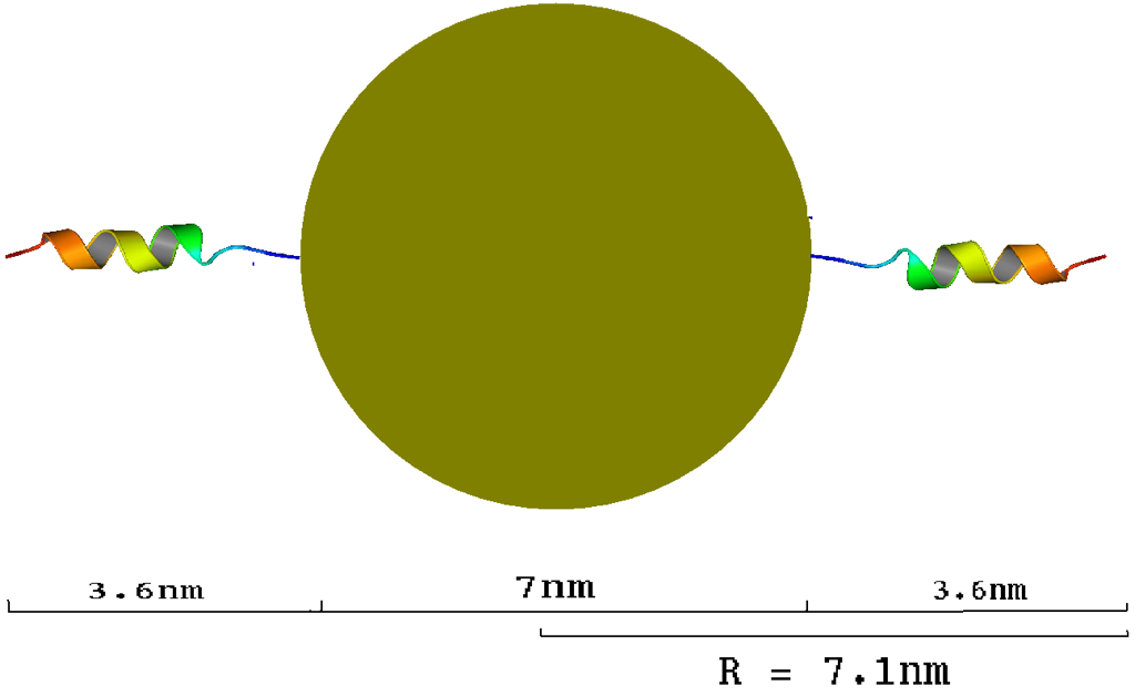
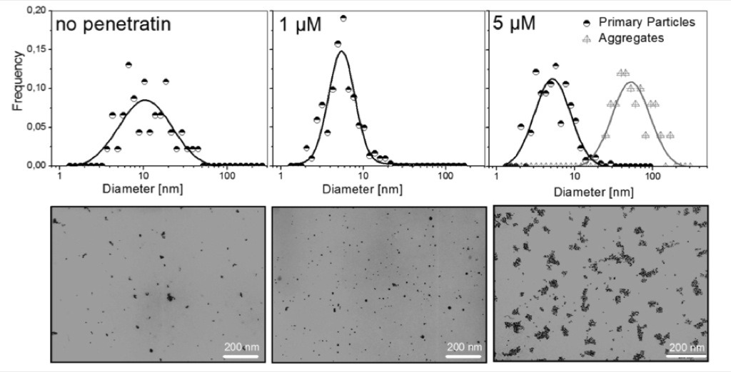 Note the dual peaks in the last histogram – these correspond neatly to the peaks we see in the DLS scans (taking into account the skewing towards larger particles that DLS is prone to). If this is penetratin we can expect there to be a maximum of 27 peptides per nanoparticle. The paper has a ton of TEM data, but no DLS data so the hydrodynamic radius of the particle is ours to infer. Based on the PDB structure of penetratin (PDB code 1OMQ) the length of an extended penetratin molecule is 3.6 nm:

so we would expect a hyrdodynamic radius of:
This is supported by comparisons done by the Barcikowski lab of TEM data and DLS data of gold nanoparticles conjugated to small DNA aptamers:
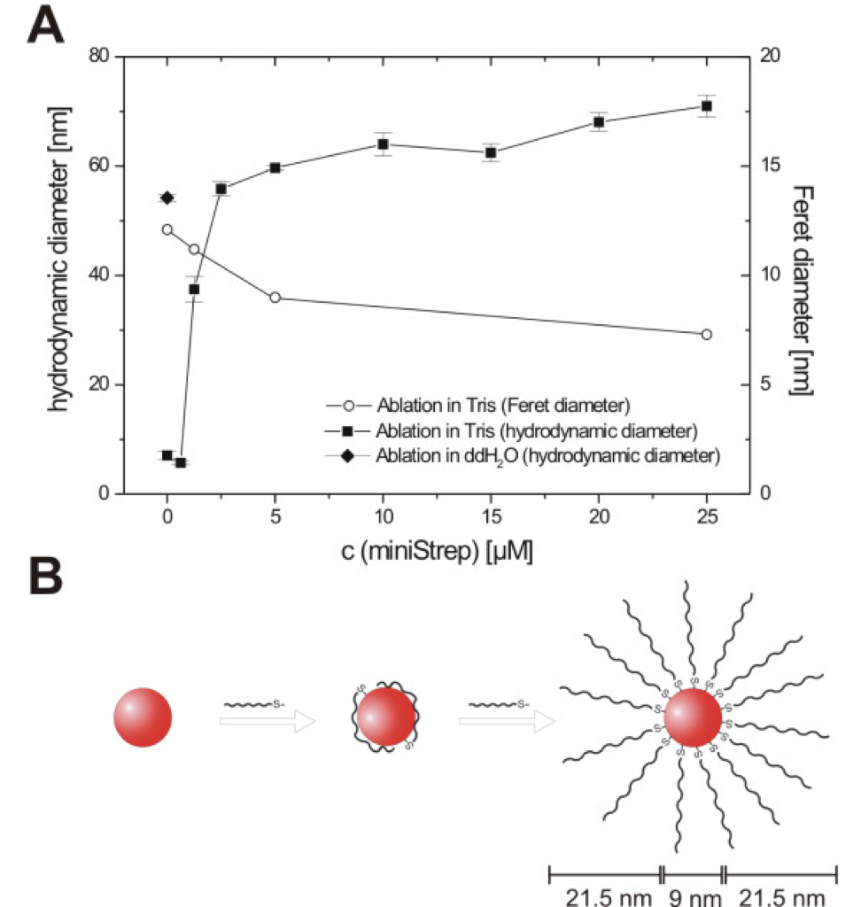
From – laser ablation-based one-step generation and bio-functionalization. It would be nice, for the ultracentrifuge step, to know the sedimentation velocity of the particles. According to the penetratin paper the surface coverage of the nanoparticle is maximal when 27 peptides are conjugated to each sphere. Since the density of gold is 19,300kg/m^3 and the molecular weight of penetratin is 2245.78, we can calculate a particle density of 2,393kg/m^3. In Stokes flow (Re < .01),

Generally, we can use this for all unconjugated gold colloids to get the following lovely graph:
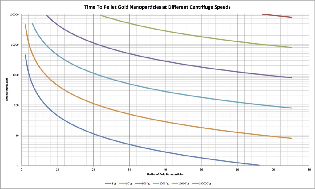
In this bioconjugated case, however, the density is an order of magnitude less, and at 10,000g we need to wait 13hr. for the colloid to travel 2cm and pellet. At 100,000g, this wait time becomes 80 minutes.
The separation of free ligand from bioconjugated particles is still a (relatively) unsolved problem, with existing chromatography and centrifugation methods of purification either damaging to the ligand or not easily scaled up. It seems like we could eek out a little paper if our Sepcons do a good job. It might be idle speculation but I envision 5 figures:
1. Ablation method and TEM histograms of particle sizes
2. Sepcon setup – if we end up using pressure, it would be something similar to: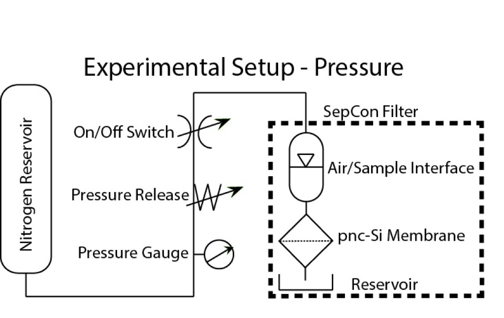
3. TECAN scans of the filtrate and retentate of successive passes of fluid through the sepcon – we should see no absorbance at 280nm in the last filtrate, and lots in the last retentate.
4. Show there is no free ligand in the retentate by spinning it down and measuring the absorbance of the supernate
5. Assay for function (we could duplicate the assay done in the penetratin paper).