Live/Dead Assay for BBB Device
A quick recap: Vaccinex has asked the lab to design a closed system device with the capability to co-culture on microporous membranes. This device would be used to mimic the blood brain barrier, with endothelial cells cultured on the apical side and glial cells cultured on the basal side, and would allow for the testing of VX15/2503, an antibody that Vaccinex has developed, in tightening the barrier.
Vaccinex provided us with two cell lines that they would potentially be using in this device: immortalized HUVECs and 2H11s. HUVECs, human umbilical vein endothelial cells, would mimic the endothelial cell layer, which form the tight junctions that prevent unwanted solutes from passing to brain tissue. 2H11s, a mouse endothelial cell line, is another option being considered for the endothelial cell layer; these cells are also used to model tumor endothelial cells [1] and could be used as a disease model. These cell lines were tested on both hybrid (C300) and MP3 (C1000) CytoVu®. Prior to seeding, each basal membrane was coated with 10 mg/mL fibronectin. After the fibronectin was removed and the CytoVu®s air-dried, each cell line was seeded in each type of CytoVu® at a concentration of 200 cells/uL. After two days of culture, the cells were imaged using a Live/Dead assay.
|
|
|
| HUVECs cultured on hybrid CytoVu® (left) and MP3 CytoVu® (right). Images are at 10X and the scale bar indicates 100 um. | |

|
|
|
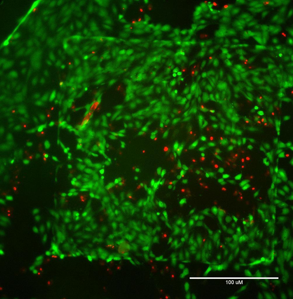
| 2H11s cultured on hybrid CytoVu® (left) and MP3 CytoVu® (right). Images are at 10X and the scale bar indicates 100 um. | |


The immortalized HUVECs grew well on both types of CytoVu® devices, with higher proliferation on the MP3 CytoVu®. The pocket of dead cells on the MP3 membrane is a little strange but could have been due human error, such as too aggressive pipetting of media. I do not think it is due to the membrane properties, because then the HUVECs would have not adhered initially or larger amounts of cell death would be visible.
The 2H11 cell line proliferated rapidly and reached confluency prior to the imaging on the MP3 CytoVu®, thus the odd shape; however, little cell death is visible, indicating no negative effects of the membrane. As this cell line does proliferate rapidly, I would suggest seeding at a lower concentration in future studies to prevent the effects seen here. There was some difficulty seeding the 2H11 cells on the hybrid CytoVu®; with the smaller basal well size, air bubbles formed during pipetting and were tricky to remove, even after multiple attempts. This is most likely the reason that few cells are visible in the hybrid CytoVu®, dead or alive, but despite these results, I do think that these cells would grow well on the hybrid device as well.
Based on the results from the Live/Dead assay for HUVECs and 2H11s on the two different types of CytoVu® devices, I believe that there will be no issue using either of these cell lines to mimic the endothelial layer in the blood brain barrier device.
[1] Yohrling-Walter, J. (2004). Clin Cancer Res 10: 2179-2189.