Diffusion Separation with w612
This was something I set up a few days ago and finally got a chance to run the gel. I was curious to try this after the previous forced separations using brain extract. If you recall, the “separation” using the pressure cell did not have a visible cutoff and all of the bands appeared in the filtrate. I figured I better try a separation with standards by diffusion to check these results. Standard diffusion setup applies here: 2ul sample, 60ul reservoir, 24 hours on teflon block. Positions are -2,2 for Trial 1 and -2,-2 for trial 2.
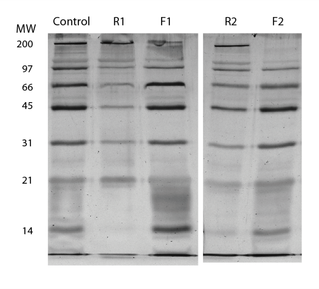
You can see that the first trial seems to have all the bands in the filtrate, although the top ones are a little lighter. The second try looks like myosin, the top band is not getting through, but it’s hard to say if maybe the first one has gotten closer to equilibrium. In both cases the lowest bands appear darker in the filtrate meaning we’re very close to equilibrium with these small species.
I think the conclusion is that we’ll need smaller pores to get convincing movable cutoffs for the paper. Unless of course that is if there’s some low density larger detect that were just not picking up by the reflection scope or the viewed area in our TEMs.
We should make note of how often it happens that we get separation with one chip but not with the next when both are thought to be pristine. This could be matter for SiMPore’s quality control division.
Is the retentate diluted to make the band intensities equivalent? If so, the second separation looks pretty good, right? Is there a link where the performance of all the nanosep, microcon, microdialysis systems with this standard ladder is provided for reference? I would like to know is this level of separation is anything to brag about…
Any idea why the cutoffs are so different? What would these gels look like if there were pinholes? Since the filtrate/retentate are different in both cases, these look like a movable separation, right? If I understand correctly, you think the pores are the same size, though, correct? Are you sure there was no difference in their handling or appearance? Were these both aggressively ozone treated (high temp, 10’s of min) to make sure they are hydrophillic with no organic surface contamination? I am confident that the silicon pores are the same size, as I have seen no evidence otherwise, however, different surface contaminents and wetting properties are possible on samples separated by an inch or two on the wafer…
Have you tried pre-exposing the membranes to 1-10 mg/mL filtered BSA first, to reduce the pore size? Since we know that we bind a monolayer of protein anyway, it is likely that your ladder itself is changing the pore size, so pre-treating with various proteins of different size may help you understand more about what is going one here. When the silanization system comes, there will be more options to test, but for now, simple adsorption seems like a reasonable approach. Try not to let the pre-treatment dry, though, and make sure you rinse in buffer, not DI – proteins bound directly to solid-state surfaces easily denature in my experience.
In response to Chris’ questions:
The retentate is diluted to the same volume as the filtrate (2ul->60ul in PBS). The second separation is what we’ve come to expect a good separation to look like. We have not performed separations using the standard ladder in nanosep and microcon due to their expense. Also comparing this experiment to nanosep and microcon is not completely reasonable as this is a diffusion only application. We have not tried standards separation in microdialysis yet. If we compare to brain extract using dialysis tubing, any movement of the lower bands in the retentate toward equilibrium is not noticed unlike in this gel (remember because of the dilution scheme reduction in band intensity of small species in retentate means they’re equilibrating).
My guess as to the difference in cutoffs is that the first trial may have been compromised by a defect. I did check these out under the scope and did not notice any pinholes. Perhaps there was something larger than the pores that I did not notice on the scope. If there are pinholes, all bands should appear in the filtrate, but I’m not sure what reduction we’d expect in the retentate (I’m gonna set up and try to image this control). These membranes had the exact same R-position on the wafer. I would expect the pores to be the same. They appeared the same and handled the same. I didn’t oxidize these samples as hydrophilicity has never been a problem in diffusion assays. This is a wet wet separation run by diffusion alone in high salts. Although if organic contamination is a potential problem, oxidation may be a route we need to take.
I’ve never tried pre-exposing the surfaces. This is also something I could test, although we are now limited by intact membranes.