Short-term Cell Adhesion Morphology
We’ve been getting some strange cell morphologies recently in short-term adhesion studies. My previous post on this subject links to a few of these results. Unfortunately, my last experiment into short-term cell morphologies on autoclaved and EtOH- sterilized samples wasn’t well-controlled since I didn’t have enough samples. Therefore, I repeated this adhesion study with proper controls. Doubly unfortunately, the backup drive on the Nikon microscope computer filled up during imaging and didn’t warn me that I was saving images onto nothing. So I lost my controls again. I wanted to post this anyway so it’s on the record. These are P9 b.End3 cells plated at 8382 cells/cm2 and allowed to adhere for 4 hours. The cells were then stained with 10uM CMFDA (in serum-supplemented media) and fixed in 4% PFA. Unless otherwise noted, all surfaces were sterilized by UV exposure overnight. I included 2 different fields of view (10X) and a higher magnification (if possible) for each surface. These are all flat (2D) surfaces, not transwells.
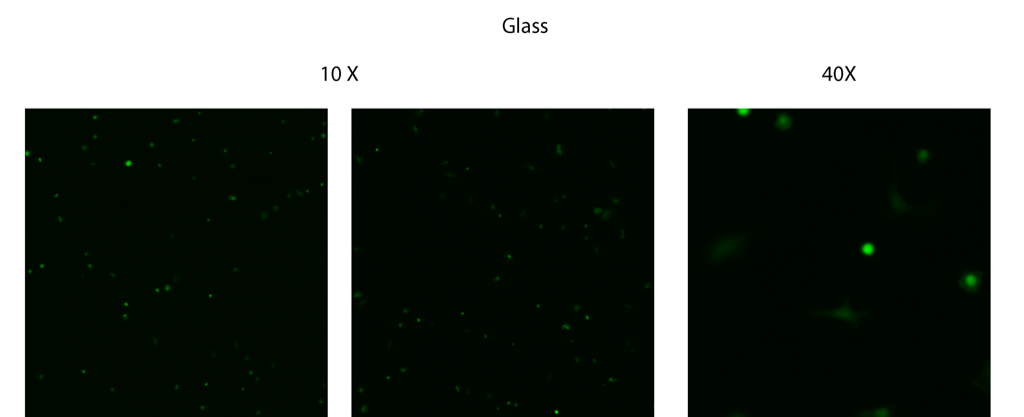
On acid-washed glass coverslips, most of the cells had started spreading or even showing a “spindly” morphology:
On acid-washed (and then PLL-coated) glass coverslips, there were fewer cells spreading than on uncoated glass:

These are cells on PTFE Teflon tape that I wrapped around a glass coverslip as a negative control. Since it’s opaque, I couldn’t get an image with the 40X objective. The cells were largely rounded with a little bit of spreading, not much different from PLL-coated glass.

On tissue culture polystyrene (TCPS), nearly all of the cells are spreading, and many of them have extended spindle-like protrusions. Of all surfaces, the cell morphology was most mature on TCPS.

On a flat pnc-Si (not a transwell) sample, most of the cells were spreading. Qualitatively, the cells look similar to those on acid-washed glass.

This is a pnc-Si sample from the same wafer (SC027) that had been RTP’ed (at 800C, 5 min, Ar) and then autoclaved. The cells are mainly rounded but spreading seems less mature than in other samples.

I lost the images of SC027 that had only been autoclaved (without RTP) and images of SC027 that had been autoclaved in the same beaker as the Sepcon plastic pieces (housing, retention ring, O-ring). Therefore, it’s hard to determing if the slightly delayed maturation/spreading in the RTP+Autoclaved sample is due to the RTP (which I suggested in my last post) or the autoclave. In any case, the morphology does not look as strange on the RTP’ed sample as in the previous post.
I also used MATLAB to do cell counting:

The blue and red bars represent the values I calculated on different surface samples (but on the same day). I only had 1 RTP+autoclaved pnc-Si sample. The glass, TCPS and pnc-Si samples showed similar adhesion to that which Anant reported for fibroblasts. Since this was the first experiment, it’s har dto say if there is a difference between RTP+Autoclaved and not autoclaved pnc-Si. Cells were poorly adherent to PLL-coated glass (not expected), which may be due to poor washing out of PLL before I plated the cells. Teflon should not promote cell adhesion, but it did. I’m not sure how to explain this.
In conclusion, it looks like there might be slight morphology differences between these surfaces, and the % adhesion values may not be significantly different. We know that cell morphology recovers on pnc-Si transwells after a couple of days and that cell growth is normal, so I’m putting this on the back burner for now. I think I have good experiment worked out now to isolate cell adhesion/morphology differences between RTP’ed, autoclaved and UV-sterilized pnc-Si chips.