Multiplexed sequential brain endothelial cell cytokine secretion measurement from μSIM
- Background
Previously, we have demonstrated multiplexed digital sensing on MCP1, KC and TNFα secreted by RAW 264.7 cells stimulated in conventional cell culturing well plates. We observed decent TNFα and strong MCP1 response but no KC secretion from RAW cells upon 100ng/ml of LPS stimulation overnight. The work verified our platform’s capability of multiplex cytokine measurements of cell secreted samples without cross reactivity among the selected analytes, secreted molecules and medium.
In our recent work, we successfully cultured mouse brain endothelial cells in μSIM and performed sequential measurement of cytokine secretion on these cells after they reach confluency and form tight junctions in the device. We only performed two timepoints (6hrs and 24hrs) in this preliminary experiment, but we successfully demonstrated we are able to utilize only 1ul of volume collected from the bottom chamber and measure the MCP1 and KC response when the brain endothelial cells are stimulated with LPS after 6hrs. In this experiment, we also try to assess the level of non-specific activation (pure endothelial cell medium (ECM) culture basal secretion) of brain endothelial cells throughout time.
- Experiments, Results and Discussions
Mouse brain endothelia cells were cultured in endothelial cell medium (ECM) in the top chamber of μSIM for ~4 days to reach confluency and form tight junctions. The devices are then separated into control (CTL) or LPS stimulated (100ng/ml) groups. Stimulants are only introduced to the top chamber of the μSIM. The devices are then incubated in a moisture-controlled cell incubator at 37 ° C. After 6 hours, 10ul of supernatant are collected from the top chamber and 2ul from the bottom chamber. 2ul of new media was replenished to the bottom chamber after the extraction. The collected samples were frozen at -80° C for later measurements and the devices were put back in the incubator. After 24 hours, all volume in both the top and bottom chamber was extracted from each device and frozen at -80° C for measurements.
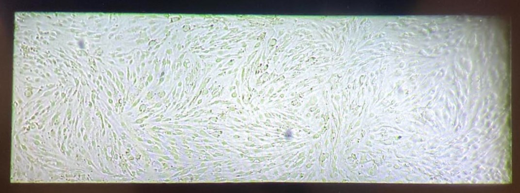 Figure 1. Representative image of brain endothelial cells cultured in the μSIM
The collected samples were thawed and diluted for digital ELISA measurements. All bottom chamber samples were diluted at a 1:30 ratio (1+29ul) and the top chamber samples were diluted at 1:4 or 1:6 ratio. All measurement results are displayed in the heat map in Figure 2. We did not observe significant TNFa secretion (<4pg/ml) from the brain endothelial cells with/without LPS stimulation. The low TNFa secretions were verified by both antibody pairs purchased through Thermo Fisher and Biolegend. MCP1 and KC secretion data were also shown in column plots for better visualization. Out of range (OOR) data shown were signals that are >1000pg/ml for MCP1 or >2000pg/ml for KC. A more aggressive dilution factor was therefore used for the second batch of measurements. Overall, we observed an increase in cytokine concentration through time in the device (24hr samples >> 6hr samples) and in general, the top chamber (stimulation source, luminal side) has a higher cytokine concentration than the bottom chamber (abluminal side). The bottom channel 6 hr incubation samples all have <2ul in volume. The raw measured cytokine concentration (after dilution) for 6hr LPS stimulated samples were 18.79pg/ml for MCP1 and 53.88pg/ml which are both well above our sensor LOD. Therefore, we should be capable of measuring cytokine secretion response in under 6hrs with the 1ul sample volume. We did observe an increase in cytokine secretion (24hr>6hr) in both chambers in the control group, indicating there might be some non-specific activation of cells throughout time or the samples collected at the 6hr timepoints were not fully diffused. Mixing and repeated experiments will be further done to verify this.
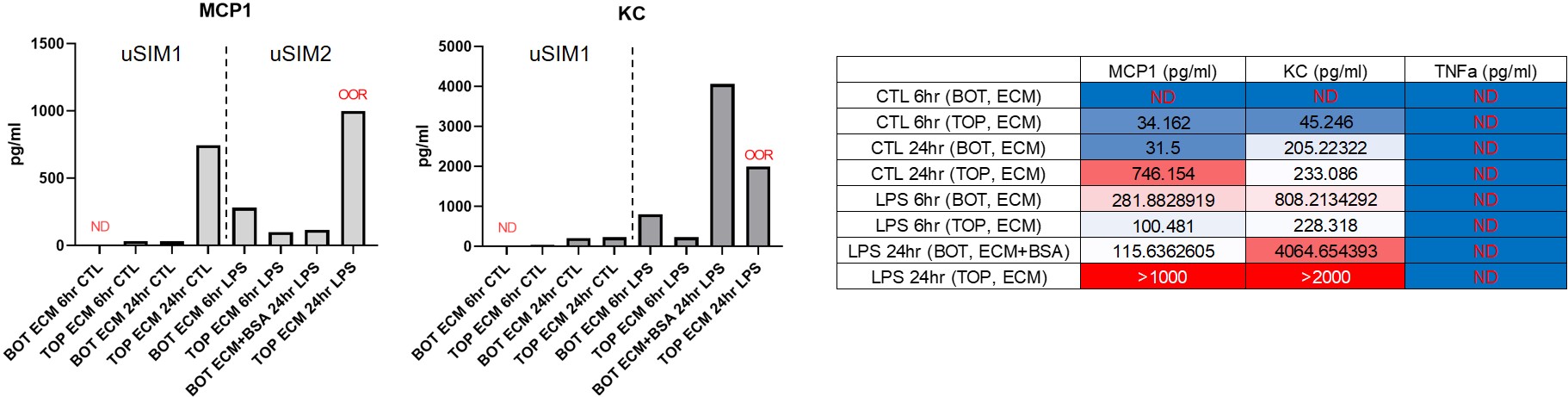
Figure 2. Heat map of all measurements and column plots for MCP1 and KC concentrations in supernatants taken out from the uSIM.
- Future Direction
We are utilizing RNA sequencing to discover the better cytokine panel to monitor from brain endothelial cells other than TNFa. We will also increase the time points taken from the uSIM for better sequential resolution in future experiments. In the meantime, we are discovering cells (e.g., microglia, astrocyte) to co-culture in the BBB system and may include different stimulants other than LPS such as serum from disease model mice. Repeats of the experiments shown above will also be done to verify consistency.