VEGF expression on pnc-Si transwell
All of my recent posts on VEGF, VEGFR2 and NF-H imunofluorescence have included data from cells grown on coverslips only. This was my first attempt to try to do immunofluorescence staining on intact pnc-Si transwells and then pop the chips out of the transwell plastic in order to image at 100X. Since VEGF is involved in angiogenesis and vacuolization, I wanted to see if VEGF expression is upregulated in cells on free-standing pnc-Si. I followed this protocol for VEGF staining but I only succeeded in popping out 1 of the 3 transwells that I had stained. These are bEnd3 cells on SC612 after 3 days of growth.
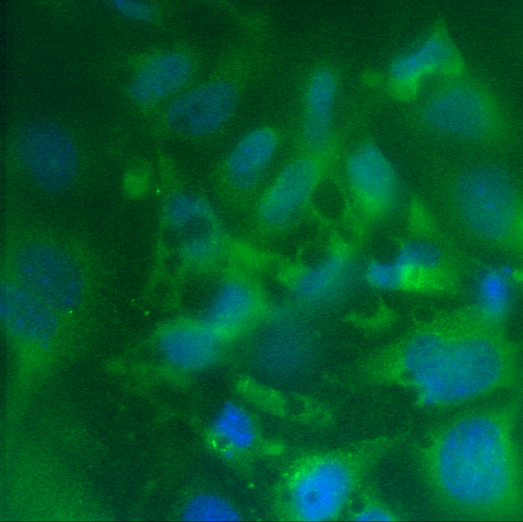
On solid pnc-Si:

On free-standing pnc-Si, you can clearly see the vacuoles – which did not stain for VEGF:
I also got some images at the edge of free-standing pnc-Si (I faintly overlaid the phase channel in these images):
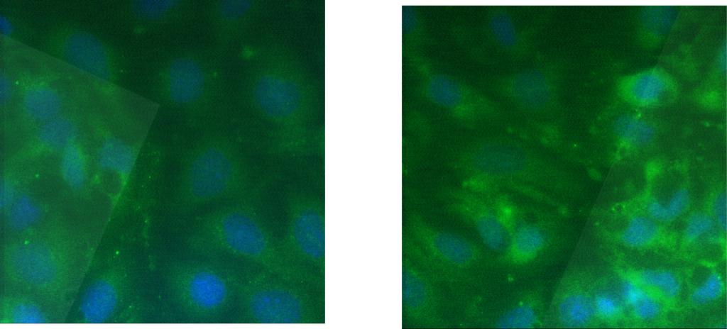
The pattern of staining on solid and free-standing pnc-Si is about the same (except for the lack of vacuole staining). I know the left image is dim but there might be some more puncta with membrane-associated cells than with cells on solid pnc-Si. Since this was only 1 sample, it’s hard to draw conclusions. I think it would be nice to have a time-course of images to see if VEGF/VEGFR2 expression changes over the 1st few days of culture.
