Optimizing Cytomix Stimulation on Mono- and Coculture BBB Models
Introduction
The µSiM BBB model is being developed at UR with the goal to understand the impact of sepsis on the brain. Here, we use an EECM-BMEC-like cell (iSPC-derived brain endothelial cells) [1] monoculture model and an EECM-BMEC-like cell and brain pericyte-like cell (BPLC; iSPC-derived brain pericytes) [2] coculture model to optimize the concentration of cytomix, equimolar mixture of TNFa + IFNg + IL1-b.
Methods
All cell culture used IMR90-4 iPSCs previously differentiated into EECM-BMEC-like cells and BPLCs or MIT-gifted healthy APOE3 (clone A20-178_CTRL-E3) differentiated into EECM-BMEC-like cells. NPSN-1L membranes were used for culture.
Monoculture
EECM-BMEC-like cells were cultured on collagen iv (Sigma C5533), bovine fibronectin (Sigma F1141)-coated devices (4:1:5 Col IV/Fn/H2O ratio) at 40,000 cells/cm2 for 6 days. On day 5 of culture, 10 – 100 pg/ml cytomix was added to the cells and cells were stimulated for 16 hr. Stock cytokine solutions are made at 100-200 µg/mL and diluted to 1 µg/mL cytomix in media first. Then two 1:100 dilutions are used to get 100 pg/mL cytomix, and finally this is diluted to the final concentration. This has provided most consistent results. Following stimulation, small molecule permeability to Lucifer yellow (LY) was analyzed. In some instances, cells were stained for ICAM-1 following the assay.
Coculture
BPLCs were cultured on the top surface of the channel of uncoated devices at 14,000 cells/cm2. They were seeded in pericyte medium, E6 + 10% FBS. The following day, EECM-BMEC-like cells were seeded in the top well of collagen iv (Sigma C5533), bovine fibronectin (Sigma F1141)-coated devices (4:1:5 Col IV/Fn/H2O ratio) at 40,000 cells/cm2 and media in both chambers was switched to EC medium, hECSR. Culture time was 7 days for BPLC, 6 days for EECM-BMECs. On day 6 BPLC / 5 EECM-BMEC of culture, 10 – 25 pg/mL cytomix was added to the cells and cells were stimulated for 16 hr. Following stimulation, small molecule permeability to Lucifer yellow (LY) was analyzed.
Results
Defining Gaps
50 and 100 pg/mL cytomix caused major cell death and large gaps. Small gaps appeared on occasion in 10 and 25 pg/mL cytomix-treated cells, and on rare occasions on non-stimulated (NS) cell layers. We believe in some cases, these gaps appear from stimulation, since it is far more common in stimulated devices, but in other cases is normal rearrangement of the cells. Sometimes it seems to lead to increased permeability, but not always. This may be due to closing of the gaps during experimentation.
Below illustrates examples of gap classifications. Permeability can be obtained for small gaps, but large gaps are disrupted barriers with permeabilities too high to plot on a chart (they overwhelm the y-axis). I have not yet defined areas for large and small gaps, but it is usually pretty clear from the picture and data when gaps are causing a fully disrupted (and non-physiologic) barrier, as opposed to just a leaky barrier.

Defining Barriers
Jim and I have talked about classifying barriers into tight, leaky, and disrupted. The utility of these classifications are yet to be determined.
Leaky barriers can occur from immature cells (i.e. low passage), improper seeding densities, and cytokine stimulation. Based on the data below and knowledge from the literature, I am thinking of setting tight barriers at Pe(LY) < 0.6 x 10⁻³ cm/min. While 0.5 x 10⁻³ cm/min is commonly used as our cutoff, other published data for IMR90-4 differentiated EECM-BMECs had transwell permeability at 0.7 x 10⁻³ cm/min, so this seemed like a good compromise, but I am open to adjustment.
Disrupted barriers are caused by large gaps that allow the small molecule to flood through and no longer enables us to determine endothelial tightness. Based on the data and images above, I think 2.0 x 10⁻³ cm/min is an appropriate cutoff.

The data above is a compilation of monoculture and coculture, which appear to be identical (at least in NPN µSiMs). I do want to note that it is nearly impossible to see gaps (large and small) in coculture devices due to the pericyte layer. It will be necessary to staining for junctional molecules post-assay.
Optimal Cytomix-Treatment and Data Analysis
Further experimentation indicated that 25 pg/mL cytomix was too high, almost always causing gaps, and often very large gaps in the monolayer (data not shown). 10 pg/mL, however, leads to a very subtle change in permeability, and the significance may be missed without properly powered experiments. I did a power analysis using the preliminary data we have, which indicates we may need as many as 22 samples per group.
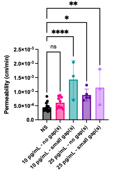
Further, I noticed that there was some variability experiment to experiment in non-stimulated devices, but the cytomix-stimulated devices always seemed to go up slightly. So, for each experiment, I averaged the permeabilities in each group and plotted the averages. Then I did a paired t-test, pairing NS and 10 pg/mL cytomix from that day’s experiment. This shows a significant increase in permeability, but may not be an appropriate analysis given the pairs are experimental days, not the same device. A power analysis indicates a paired test will only need 6 samples per group.

Conclusions
Cytomix stimulation of 10 pg/mL appears to be the best tolerated concentration for both IMR90-4 and A20-178_CTRL-E3 EECM-BMEC-like cells and IMR90-4 coculture of EECM-BMEC-like cells and BPLCs. The treatment results in only occasional gaps that are usually small and lead to leaky, not disrupted, monolayers, which can be categorized as needed. However, the permeability changes are subtle, so experiments need to be properly designed. Alternatively, 15 and 20 pg/mL cytomix could be tested.
Other Data

ICAM-1 staining is another indication that cells are responsive to 10 pg/mL cytomix, even when permeability changes are small.
References
[1] Nishihara, H., Gastfriend, B.D., Kasap, P., Palecek, S.P., Shusta, E.V., and Engelhardt, B. (2021). Differentiation of human pluripotent stem cells to brain microvascular endothelial cell-like cells suitable to study immune cell interactions. STAR Protoc 2(2), 100563. doi: 10.1016/j.xpro.2021.100563.
[2] Stebbins, M.J., Gastfriend, B.D., Canfield, S.G., Lee, M.S., Richards, D., Faubion, M.G., et al. (2019). Human pluripotent stem cell-derived brain pericyte-like cells induce blood-brain barrier properties. Sci Adv 5(3), eaau7375. doi: 10.1126/sciadv.aau7375.