S1P Receptor mRNA Quantification in HUVECs and HPMECs (EC Heterogeneity)
Introduction
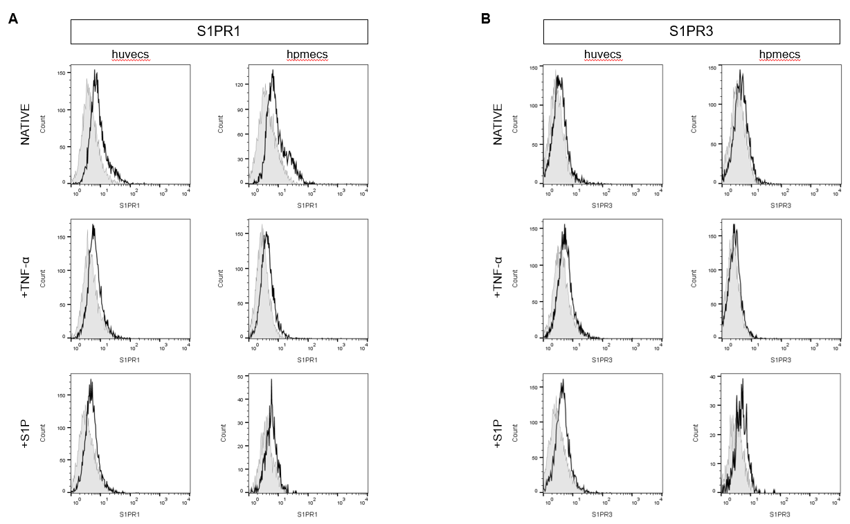
Sphingosine-1-phosphate (S1P) is a bioactive sphingolipid with known paradoxical effects on endothelial cell (EC) barrier regulation. S1P binds 5 GPCRs: S1PR1 (Endothelial differentiation gene (EDG)-1), S1PR2 (EDG-5), S1PR3 (EDG-3), S1PR4 (EDG-6), and S1PR5 (EDG-8). Despite the aliases, endothelial cells predominantly express S1PR1&3. S1PR1 has been shown to have barrier protective or enhancing effects on ECs, while S1PR3 is hypothesized to trigger thrombin-like barrier disruption. Previously, we explored the effects of S1P on native EC barrier (as measured by TEER). We found that S1P was barrier protective in HUVECs and barrier neutral/disruptive in HPMECs. To further understand this result, we characterized surface expression of S1PR1&3 on both HUVECs and HPMECs with flow cytometry. We found similar population levels of S1PR expression (e.g. S1PR1 on HUVECs was similar to S1PR1 on HPMECs). Interestingly, dot plots showed higher levels of HPMECs predominantly expressing S1PR3 as compared to HUVECs. Thus, we concluded that HPMECs are likely to exhibit barrier disruption upon S1P stimulation due to a subset of cells expressing S1PR3, opening the whole monolayer up to barrier breakdown. Understanding this EC heterogeneity (both on a single cell and cell line level) may be key to S1P therapeutics with refined effects. In a follow-up to this work, we wanted to characterize S1PR mRNA in the two cell lines.
Relevant Blog Posts
Introduction to S1P & Initial TEER Data
S1PR Surface Expression w/ Flow Cytometry
Methods
Quantitative Reverse Transcription Polymerase Chain Reaction (RT-qPCR)
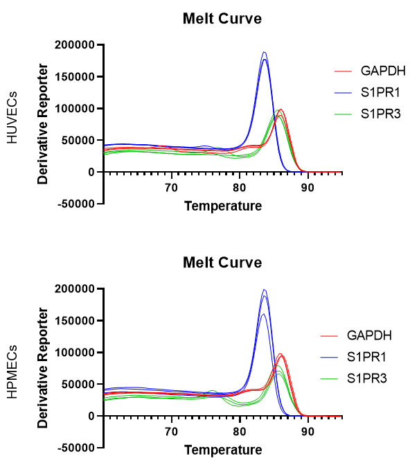
RNA was isolated from HUVECs or HPMECs using a Qiagen RNeasy plus mini kit, either following trypsinization and pelleting, or directly from the culture well (lysis buffer added to washed cells, cells removed with bent p200 pipette tip). In select cases, ECs were pre-stimulated with TNF-α [20 ng/ml] for 24 hours prior to RNA isolation. Once isolated and quantified, RNA was converted to cDNA using the Invitrogen SuperScript™ III First-Strand Synthesis System for RT-PCR. ThermoFisher Power SYBR Green Master Mix was used for qPCR. The following primers were obtained from IDT: 5′-ACCACAGTTCATGCCATCAC-3′ and 5′-TCCACCACCCTGTTGCTGTA-3′ (GAPDH), 5′-GACTCTGCTGG CAAATTCAAGCGAC-3′ and 5′-ACCCTTCCCAGTGCATTGTTCACAG-3′ (S1PR1), 5′-CAAAATGAGGCCT TACGACGCCA-3′ and 5′-TCCCATTCTGAAGTGCTGCGTTC-3′ (S1PR3) [Lin, C. I., Chen, C. N., Lin, P. W., & Lee, H. (2007). Sphingosine 1-phosphate regulates inflammation-related genes in human endothelial cells through S1P1 and S1P3. Biochem Biophys Res Commun, 355(4), 895-901. doi:10.1016/j.bbrc.2007.02.043]. GAPDH was used as an internal control. The following qPCR controls were run in combination to the standard template + primer technical triplicates: No enzyme RT-PCR control for all genes and no template (water) control for GAPDH. Statistical analysis was performed in graphpad prism.
Results
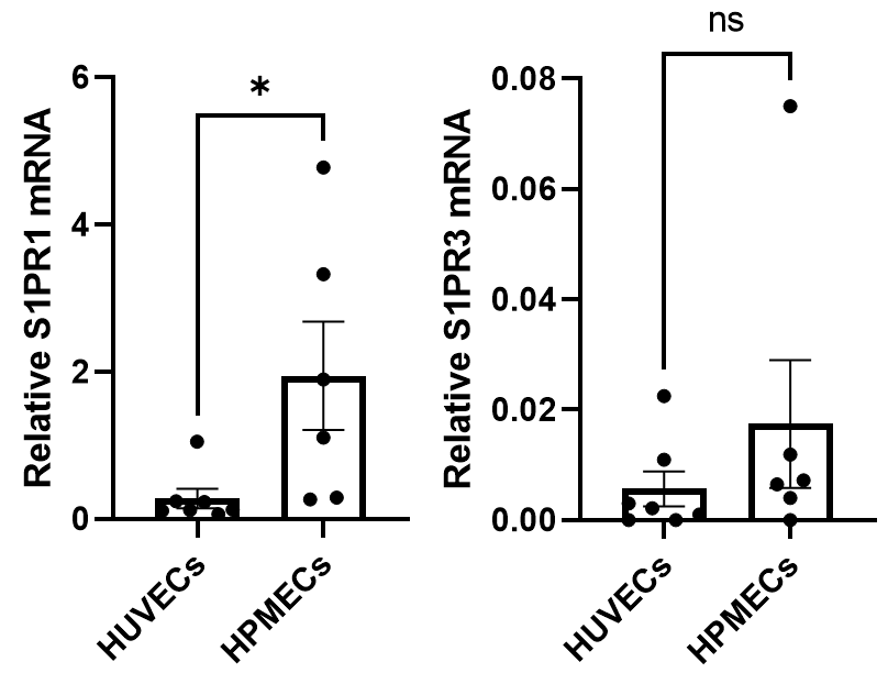
S1PR1 and 3 mRNA were quantified in HUVECs (Figure 1) and HPMECs (Figure 3). Both cell lines express significantly more S1PR1 mRNA compared to S1PR3 mRNA. Statistical comparison across cell lines showed significantly more S1PR1 mRNA for HPMECs as compared to HUVECs (Figure 3). No significance was detect across cell lines for S1PR3.


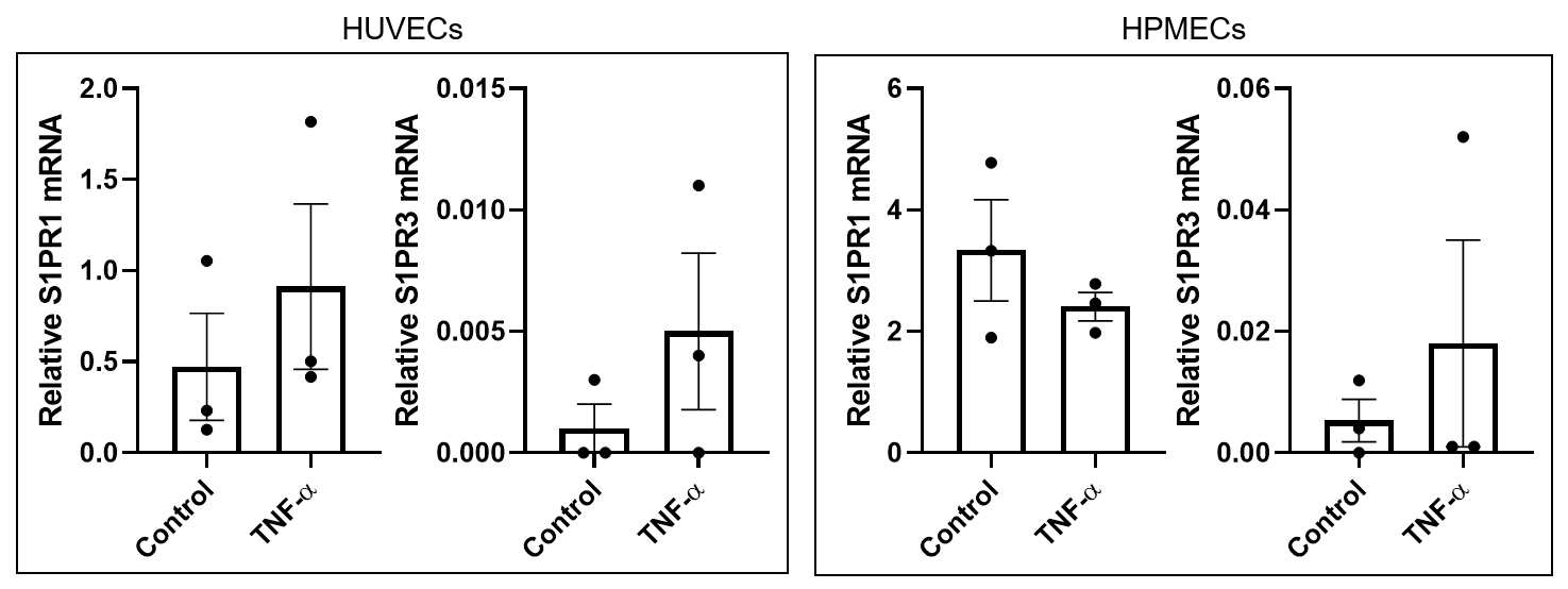
The ultimate goal of this work is to better characterize the heterogeneous effects of EC lines to S1P in an effort to guide therapeutic development for the treatment of severe inflammatory disease. Thus, we wanted to recharacterize the receptor mRNA in these cells under inflammatory conditions. In this case, we saw trends in increased receptor (both S1PR1 and 3) mRNA in HUVECs. Neither receptor exhibited statistically relevant changes in mRNA levels in HPMECs following TNF-α stimulation.
Conclusions
RT-qPCR was relatively successful on these cells and genes. RNA yield is lower for HPMECs compared to HUVECs in general, but enough template is isolated for RT and qPCR as defined in the respective protocols. Interestingly, HPMECs appear to express higher levels of S1PR1 at baseline, which is in slight contradiction of our previous TEER results (based on the notion that S1PR1 is barrier protective). Relating these data to the function output is less informative compared to surface expression of the receptors, however. Future work will continue to utilize flow cytometry to understand S1PR heterogeneity on these two EC lines. This will include repeating the previously presented flow work following TNF-α and S1P stimulation of ECs. Repeating TEER work on TNF-α stimulated ECs is also in the timeline for this work, as this was previously performed on unstimulated ECs only. Final experiments will explore the consequence of S1P stimulation on neutrophil migration.
Update