Collagen Uni-axial motion Introduction
This post serves as an introduction to the uni-axial motion envisioned to be integrated into the tendon-on-chip platform. As discussed previously we are working in junction with Dr. Awad from the Center for Musculoskeletal Research (CMSR) center at UR to develop an in vitro platform to study fibrosis and inflammation in tendon tissue. One important aspect of this platform is the ability to induce a stretching motion in the tissue to analyze its effect during tendon healing.
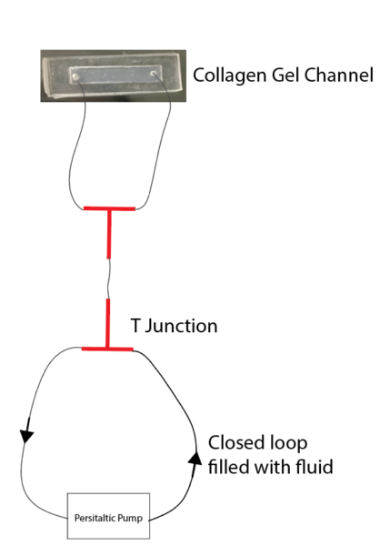
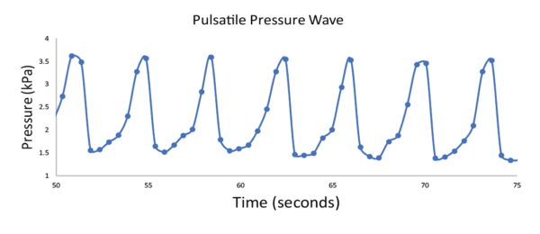
Our attempt to simulate this uni-axial motion in our platform takes advantage of the pressure changes produced during peristaltic motion. By using a peristaltic pump and tapping into this pressure difference we are able to translate motion into the type I collagen gel.
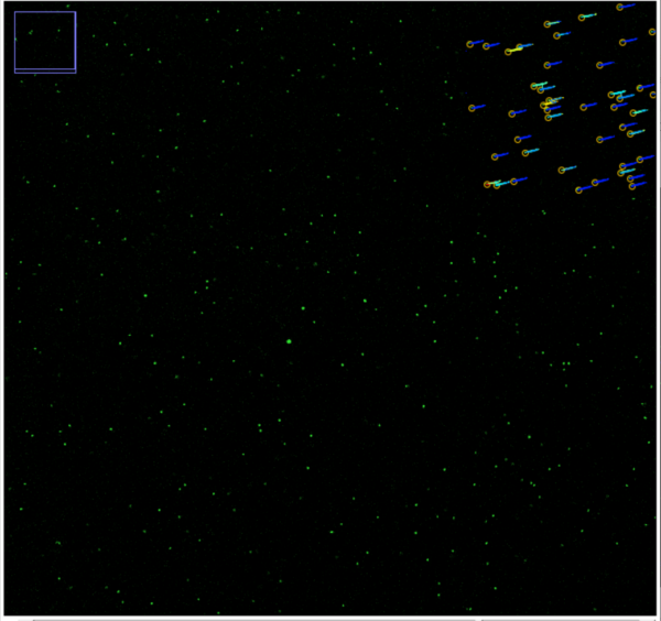
We analyzed the translation of this motion into the collagen gel using the dragonfly. I have performed the imaging on two gels. This first time I only analyzed the center of the gel, and the second time I analyzed the edges and the center. From the scope, we can define the location and obtain a precise x,y,z location for each imaging section.
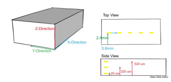
This first image references the locations in 3D space that are analyzed. The collagen gel channel is 25mm long, 5mm wide and 750 um tall.

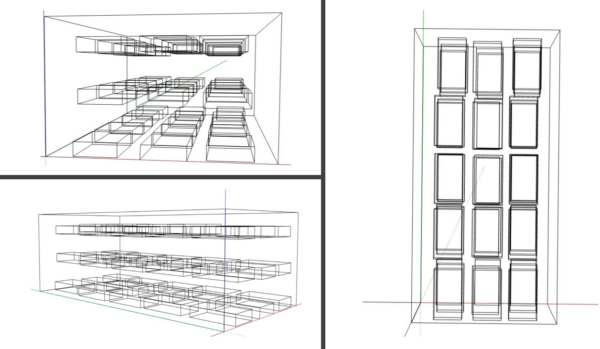
From this analysis, we obtain an image series at each location.
The next step is to perform particle tracking to obtain strain information from this movement. The strain we are aiming for is 1-5% as this is what is expected in vivo. Some MATLAB programs are available to perform this analysis including Digital_Image_Correlation, and PTVLab. I am not well versed in this aspect, yet I have made some progress in obtaining engineering strains for different sections.
This is a strain for a collagen section at the bottom of the gel (Close to the glass slide) where there was low movement observed.
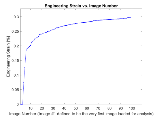
This is the strain observed for a section in the center where there is a higher movement observed.

From this initial analysis, I noticed there are drastic differences in the locations of the collagen gel with the highest strains observed in the center of the gel. Although there does not need to be identical strains in the gel for the experimental analysis, it will be necessary to obtain a more complete strain map for the behavior of the gel. With this information, we can have a better translation into a cell-seeded hydrogel.

The next steps include moving onto BlueHive to perform post-processing and strain analysis.
To be continued…..