Preliminary Testing for Transwell Cancer Exosome Co-culture Studies
Last time this project was discussed at NRG, Henry was the project lead (we miss you, Henry). He was observing bladder epithelial cell (BdEC) invasion and migration in co-culture with T24 bladder cancer cells. Here are links to his previous posts.
https://trace-bmps.org/bdec-t24-co-culture-wound-healing/
https://trace-bmps.org/bdec-t24-co-culture-invasion-assay-preliminary/
We have transitioned away from the invasion assay and moved towards transwell migration. In the future, we would like to perform a co-culture between the two cell types with the transwell assay to determine if a life-like concentration of cancer extracellular vesicles (EVs) causes increased BdEC migratory behavior. For now, we are completing preliminary testing for these experiments.
1. Parylene membranes vs standard track-etched membranes


For co-culture experiments, we will be comparing migration on Thincert track-etched membranes to parylene membranes. To better match the porosity of the track-etched membranes, we will be using parylene membranes with 10% porosity.
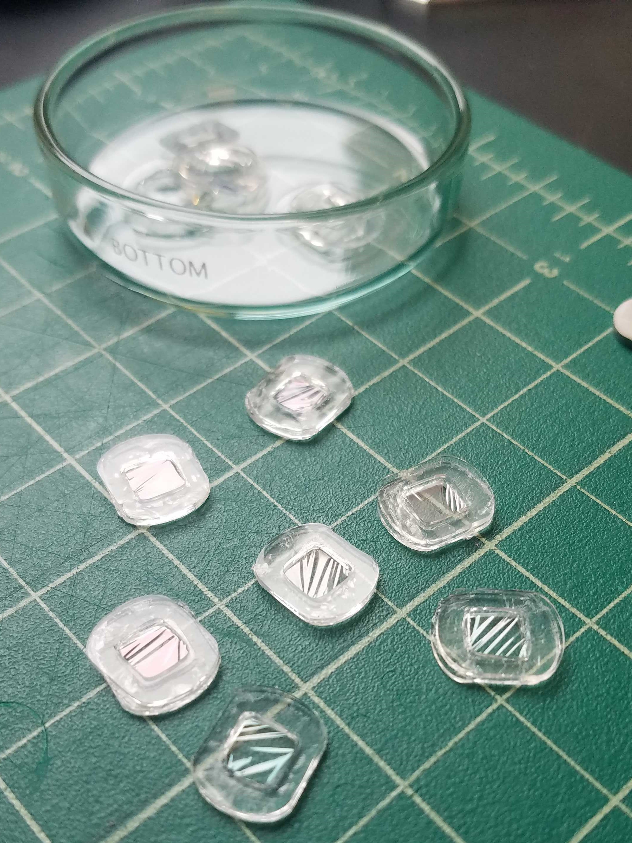
2. Fabrication of parylene transwells
- Track-etched membrane removed from Greiner BioOne Thincert.
- Pressure sensitive adhesive added to outer edge of transwell.
- Transwell with adhesive attached to wafer.
- Transwell placed in DI water until membrane separates from wafer.

3. Fixing and imaging cells on both sides of the membrane
We plan to count cells on both sides of the membrane during co-culture experiments to determine how many cells have transmigrated. This will be accomplished by trypsanizing the cells and counting using a hemocytometer. Additonally, SEM images will be taken of some transwells in order to determine if our counts are accurate. We need to find the best method of fixing our cells on these membranes in order to take SEM images. We are testing the following methods:
- 4% Paraformaldehyde
- 4% Glutaraldehyde
- 4% Formaldehyde – cold
- 4% Formaldehyde – warm
- 2% Glutaraldehyde & 4% Formaldehyde
- 2% Glutaraldehyde & 4% Paraformaldehyde
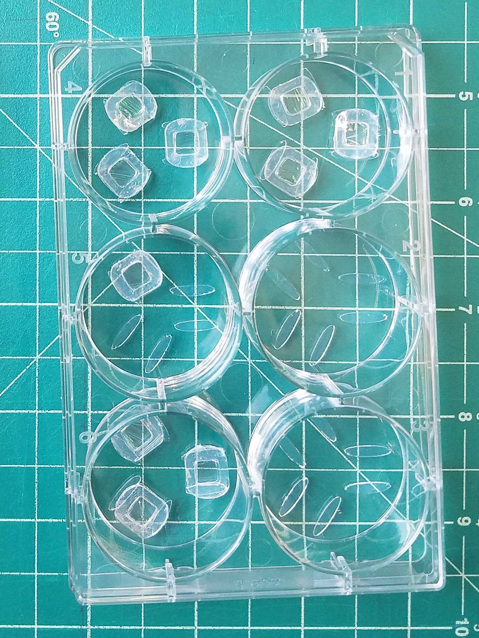
T24 cancer cells will be seeded onto parylene membranes using the devices shown below. Samples will be given a 24h incubation time after attachment to allow for transmigration through the pores. Cells will then be treated with one of the above conditions. Following this, the wells will undergo a series of ethanol treatments to gradually dehydrate the samples. The last step includes immersing the samples in 1mL of 100% ethanol and leaving them to dry overnight.. The following day, the devices are able to be imaged using SEM. The whole process for fixing cells is outlined below.
1x PBS –> treatment according to list above –> 1x PBS –> 25% ethanol –> 50% ethanol –> 70% ethanol –> 90% ethanol –> 100% ethanol (x2)
We are currently running this experiment and will take images on Thursday. The best condition will be used for future fixation for co-culture studies.
The best way to add more stability to these membranes is to add SU8 frames to these membranes, based on the transparency mask that we made for this purpose:
https://drive.google.com/open?id=1Pa5A0xyN7qPXQvicXR_24xTtJDNiPz8c