Lung on a Chip
Links and Papers:
Lung on a Chip Application (Pulmonary Edema)
Chris came across the publicity video last week and Jim asked me to look into the specifics. It’s a pretty nifty device.
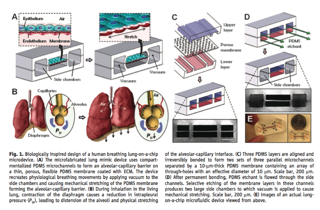
As the video shows, the device is a co-culture structure fabricated entirely from PDMS. The membrane is 10  thick. The channels are on the order of 100 high by 200-400 wide. They use mortar bonding to join top, filter, and bottom layers, with a thin layer of PDMS. There are two adjacent vacuum chambers that can compress or stretch the membrane to simulate physiological stress during breathing. In order to make the vacuum chamber, an etchant dissolves the filter in those chambers to merge the top and bottom layers. Epithelial and endothelial cells are grown on the membrane, and then conditioned by the stretching/relaxing membrane.
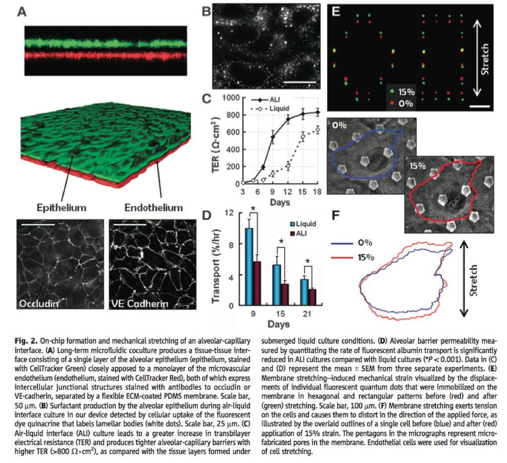
The amount of stretching is an additional 10-15%. The pnc-Si membranes may not be an optimal membrane for this application. The authors track cellular movements across the barrier through the large pentagonal holes in the membrane using fluorescence and phase microscopy. They assess the cell barrier with TEER, but don’t provide any detail about their measurement scheme. Fluorescent albumin is used to quantify some transport too. Ultimately, the authors wanted to create a realistic model for pulmonary edema (simplistically, excess fluid buildup on the alveoli); current in vitro models only produce a minimal amount of fluid. The authors hypothesize that the mechanical stretching of the lung would exacerbate the underlying problem. They recovered about 3x times more fluid in their device compared to another in vitro model and then compared to a mouse lung model through a permeability study.

Don Ingber’s group really does research in diverse areas. They also came up with a microfluidic device where they used immuno-magnetic beads that were used as magnetic opsonins to separate fungi from blood, as a strategy to fight sepsis!