RNA extraction from captured exosomes
As a proof of principal experiment, we wanted to test if Kilean and I could extract RNA from exosomes following their capture on 1298 membranes. As noted in Figure 1, we first used ultrafiltration to capture exosomes on the nanoporous membrane. We then attempted exosome lysis and RNA release by flowing the lysate below the membrane. The presumption is that the tangentially flowing lysis buffer (containing guanidine-isothiocyanate and β-mercaptoethanol) will flow up into the underside of the pores, lysing the captures exosomes and releasing their RNA. This lysate would then flow to the outflow channel and be collected. RNA was then extracted using silica-based affinity columns from a commercial kit (RNeasy Qiagen) and levels of 3 housekeeping gene transcripts was determined by RT-qPCR. To assess the efficiency of release of captured exosomes RNA we compared the captured exosome’s housekeeping gene RNA levels with that from directly lysed exosomes.
For capture we flowed ~200 ml of Hansa exosomes (at 1 x 10⁸ exosomes /per ml) at 10 μl/min with 4 μl/min pull. Lysate had been preloaded into a tube (500 ml volume) attached to the input port of the lower chamber (see Figure 1) but had been clamped off during the initial capture step to prevent premature lysis. Following the capture step, the clamp was removed which permitted the lysate to be pulled into the device. The transmembrane pressure (and exosome flow into the chamber) was maintained until the remaining ~800 μl of exosomes had flown into the device. Two observations at this point, first, b-mercaptoethanol (highly volatile and readily detected by odor) was not “detected” by odor in the out flow from the upper chamber, indicating that significant movement of lysate from the lower the upper chamber did not occur. Second, the total volume of recovered lysate was almost equal to the input volume. Together these two finding suggest that following capture and initial exposure to lysate, minimal upper chamber fluid (and possibly exosomes) moved into the lower chamber. This hypothesis needs to be tested, but if true may indicate that the trapped. Lysed exosomes clog the pores.
In order to assess the presence of released exosomal RNA we examined the level of 3 housekeeping genes (fragments of which can be found in bulk exosome preparations) by RT-qPCR. As an estimate of RNA collection with the device (a mixture of efficiencies of capture, lysis, purification, reverse transcription and qPCR) we compared the collected “exosome” lysate to 200 μl of exosomes that were directly lysed in the lysis buffer. Note that this is the volume of exosomes flowed through for capture but 1/5th the total volume that went into the device. Figure 2 shows the ratio of the housekeeping gene RNAs in captured to direct lysed exosomes (in 3 capture experiments). These values range between 0.8 to 1 indicating that the captured exosomes have between 80 and 100% of the RNA of a 200 μl exosome sample. In future experiments we will: 1. examine the kinetics of exosome lysis. 2. determine if the lysis treatment of captured exosomes clogs pores. 3. Determine the fraction of captured exosomes that is lysed. 4. Examine if simultaneous running of exosomes in the upper chamber and lysis buffer modulates the yield of RNA capture.
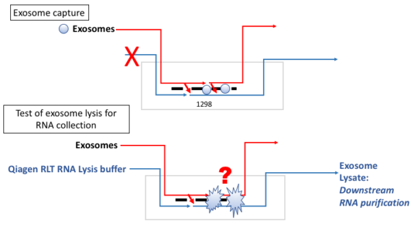
Figure 1. Model of exosome capture and RNA lysis.

Figure 2. Ratio of captured to direct lyses exosomal RNA. RT-qPCR was performed on samples from the lower chamber outflow channel (‘captured’) and unprocessed exosomes (‘direct lysed’) using three housekeeping gene targets. The ratio of captured to direct lysis RNA was determined for 3 separate capture experiments. Error bars are standard deviation.