BMES 2017
Here is a summary of some of the talks I attended at BMES 2017, mainly from the Nano and Micro- technologies track. This is an ongoing post, I will post more comments, links, and figures.
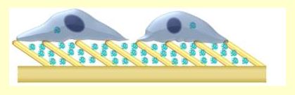
- Anna Kalmykov (Tzahi Cohen-Karni) Carnegie Mellon University: 3D Nanosensors for Electrical Interrogations of Engineered Micro-tissues. Overall synthesis of nanostructures to be used as electrodes in biosensors. Their fabrication process uses micellar nanolithography to grow and place the array of Au nanoparticles. The schematic below shows their “fuzzy graphene” which grows from Si nanopillars, this materials was not part of their talk but it is interesting (here is the paper).

- Amy Mantz (Angela K. Pannier) University of Nebraska-Lincoln: Sculptured Thin Films Alter Cellular Actin Features and Transfection Efficacy. Use of a nanostructured cell culture substrate containing Ti nanopillars for DNA storage and transfection. The cells (NIH 3T3) showed different morphology when grown on nanopillars of different length. For flat substrates and long nanopillars (100 nm) the cells developed long filopodia (attributed to the lack of features, also mimicked by long features). For medium length nanopillars (50 nm), the cells showed membrane ruffles and transfection efficacy was increased. Effect was lost for smaller nanopillars (25 nm).
- Alec Smith (Deok-Ho Kim) University of Washington: Nanopatterned Conductive PEG/Graphene Hybrid Scaffolds for Cardiac Tissue Engineering. Conductive scaffold for improved phenotype of cardiac cells guided by conductive topographical cues (paper here).

- Geonhui Lee (Dong-Hwee Kim) Korea University: Networked Concave Microwell Arrays for Constructing 3D Cell Spheroids. The material included in the talk has not been published. The basic idea is a series of microwells that are connected through a bottom membrane as well as by side wall channels to neighboring wells. The “membrane” is a really thin layer or Si, leftover after etching the wells. His website shows a long list of interesting papers comprising microwell arrays,tissue engineering and exosomes, all marked as “Manuscript under consideration”.
- Christopher Chapman (Erkin Seker) University of California: Nanoporous Gold Biointerfaces: Modifying Nanostructure to Control Neural Cell Coverage and Enhance Electrophysiological Recording Performance
- Zidong Li (Erkin Seker) University of California: Electrically-Controlled Small Molecule Delivery from Nanoporous Gold Electrode
- Jovana Veselinovic (Erkin Seker) University of California: Electrically-Guided DNA Printing and Multiplexed DNA Detection with Nanoporous Gold Electrodes in Microfluidic Devices
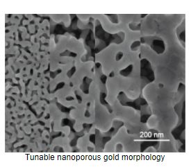
- Christopher Chapman (Erkin Seker) University of California: Nanostructure Introduces Artifacts in Quantitative Immunofluorescence by Influencing Fluorophore Intensity. This group presented their nanoporous Au, which is fabricated form desorption of Ag from a Au-Ag alloy. The morphology (pore size) can be controlled by tuning the thermal treatment used for Ag desorption. The resulting nanoporous gold is used mainly as electrodes for brain tissue biosensors. The porosity increases the surface area of the electrode and decreases impedance by blocking larger molecules from adhering to the electrode (acting as a filter).

- Praveesuda Michael (Steven G. Wise) Th Heart Research Institute, Australia: Novel Nanocarriers Capable of Spontaneous, Linker-Free Multifunctionalization. One step fabrication of nanoparticles.
- Walter Varhue (Nathan Swami) University of Virginia: Deformability-based Separation of Pancreatic Islets from Exocrine Acinar Tissue for Transplant Applications
- Nari Hong (Yoonkey Nam) KAIST, Korea: Manipulation of In Vitro Neurite Outgrowth Using Nanoplasmonic Neural Interface Platform
- Shang Song (Shuvo Roy) University of California: A Silicon Nanopore Membrane Based Intravascular Bioartificial Pancreas Device for Islets Encapsulation Under Convective Transport
- Po-Hsun Huang (Tony Jun Huang) Duke University: Delivery of Undiluted Whole Blood in Microchannels Enabled by Acoustic-based Fluid Propulsion
https://arxiv.org/pdf/1402.4152.pdf
http://www.pnas.org/content/112/16/4970.full.pdf
http://www.pnas.org/content/112/16/4970.full.pdf