Sizing of ultrasound contrast agent and sub-harmonic imaging
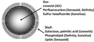
Contrast agents are used to increase received signal in imaging systems. In the case of ultrasound, such agents are small micron sized gas bubbles. Ultrasound measures the change in acoustic impedance (product of density and speed of sound), but since the speed of sound in tissue is relatively constant, medical ultrasound effectively measures changes in density. Now, the reason a gas bubble acts as a contrast enhancing agent is because at its boundary there is both a change in the speed of sound and density of the medium, making the acoustic mismatch extremely high, essentially amplifying its signals signals with increased reflection. The gas shell however makes these bubbles unstable, so it is now common practice to add some sort of lipid shell (proteins as well) to create a longer lasting agent (the core gas is also generally a fluorine complex to decrease passive diffusion). The increased stability of these bubbles has allowed applications to move from contrast improvement to other analytical techniques, assuming they are below 10 microns in diameter so not to be filtered by the lungs.
In addition to the issue of lung filtration, small size also allows better control of non-linear effects and transport to micro-vasculature in tumors. The bubble acts as an oscillator in the field, and can thus be assigned a resonant frequency (another consideration is the frequency of imaging, which drives resolution). Sonication at this frequency creates a dramatic increase in micro-bubble signal, and can also generate higher harmonics, that is frequency multiples of the resonant frequency. As these signals are unique to micro-bubbles they offer the chance for super-resolution imaging and other exploitable phenomena. The one I am most concerned about in my research is the sub-harmonic (f0/2 in the frequency domain).
The subharmonic signal from micro-bubbles is a thresholded phenomena, depending on the ambient pressure of the environment. Physically, the sub-harmonic is generated when the incoming pressure wave causes the bubble to collapse in on itself, or buckle. A major goal of my research is to use the overall sub-harmonic signal to determine the ambient pressure. Currently, this is done by performing an incision in the femoral artery and feeding a pressure catheter to the sight. It only takes 40Pa of pressure to damage a lumen wall, so in addition to being incredibly invasive, the procedure will likely create more issues than it would solve.
The issue now is that in order to get accurate pressure readings from the sub-harmonic signal, we need it to have exceptional signal to noise and contrast to noise ratios. Some efforts have been focused on adjusting the emitted pressure signal to enhance bubble activation, but these methods are ultimately limited by the quality of the distribution. A much more effective approach to improving signal is to adjust the distributions themselves. Direct fabrication of mono-disperse bubbles has been achieved using micro-fluidic devices and other techniques, but the manufacturing process has either not been cleared for use in humans, restricting it to pre-clinical work, or cannot create concentrations high enough for imaging (10^9 bubbles/ml). One specific example consists of work done by my predecessor, Himanshu Shekar. He was able to show that bubble size modification could be achieved using a centrifuge and floatation techniques, as well as an improvement in harmonic signal, shown below:

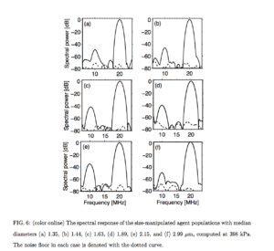
Now part of the difference between sizes in this case is also do to the transmitted frequency. Since it was 20MHz throughout, the bubbles whose resonant frequency was that of the transducer (2micron here) will have a better signal, but the additional improvement from the noise floor is a result of the more coherent signal from this distribution. Even though this method yields appropriately distributed bubbles, it is not rigorous enough for use in clinical and medical settings.
My goal is to overcome this difficulty using the microchip filtration devices used by the McGrath Lab. Using these chips, we should be able to make the distributions even narrower, making the non-linear response even greater, as well as controlling the pressure threshold of the sub-harmonic. Without a coulter-counter, sizing of these particles can be difficult as their stiffnesses is a function of their radius, the parameter we are trying to isolate. My hope is to combine measurements from the zetasizer once it is fixed with microscope images and a flow model to ensure the most accurate sizing and highest sub-harmonic response. In addition, I am also currently working on a Comsol model of 3d flow across the chip, with the hope of adding the bubbly flow module to completely simulate my environment.
For your COMSOL model, what is your justification for using bubbly flow? I believe that bubbly flow is for systems that have two phase flow, a liquid phase and a gas phase. This assumes that the second phase is something like air or hydrogen which behaves as a compressible fluid, while the first phase, which is water or media or buffer is an incompressible fluid. With the second phase as a compressible fluid and the first phase as an incompressible fluid, then you are justified using the bubbly flow system, because the compressibility of the second phase significantly affects the flow profile. For your system, yes you have these “gas” bubbles, but they are coated and essentially behave like solid particles. So in this case, you would be modeling particles in single phase flow as the particles that you are capturing are not compressible (or at least I really hope that they aren’t).