Manipulating neutrophil migration through the vascular barriers
For last many months, I am working on neutrophil migration experiments to develop protocols to inhibit them from traversing the basement membrane. My previous blogs are here and here. I want to prevent neutrophils from crossing the endothelial basement membrane (BM), and record the changes in TEER and try to correlate with the gain in permeability during infection. I plan on using the antibody against beta-1 integrin to inhibit neutrophil-BM migration since they are the main receptors for all the ligands found in ECM.

As you can see, variety of subtypes of beta1 integrin bind to ECM proteins. BM is dominated by laminin and collagen (4), so blocking the activity of Beta1 is hypothesized to reduce, if not eliminate the migration of neutrophils on basement membrane.
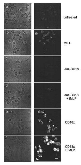
I am using this antibody to block beta 1 integrins on human cells. Now here is the catch: human neutrophils have very few copies of beta 1 integrin on their surface during inactive or resting state. Their upregulation happens only when they get activated, or when their beta2 (CD18) integrins get activated, as proved in this paper.

This also makes sense; when neutrophils get marginated in the microvessel for transmigration, they first need the selectins and then the beta 2 integrins for crossing the endothelial barriers. It’s only when they cross the ECs, that they will need the next set of integrins to crawl on the ECM proteins. So pre-labeling with anti-beta 1 outside the chamber when neutrophils are not activated is not going to be of much use. I need to label neutrophils in situ when neutrophils are ligated to the endothelial ligands and activated from fMLP from the basal compartment.
So I isolated neutrophils fresh from my own blood and used for my experiments. HUVECs were grown on 100-micron type 1 collagen on 3-micron flat glass membrane, with 600 microns of collagen gel below the chip in the basal chamber. I added neutrophils in MCDB media with enough antibody (1 ug per 1 million of cells in 100 ul vial) in the top chamber, and 10 nM fMLP in the basal chamber right after neutrophil addition. After 3 hours of incubation, I counted the cells transmigrated and divided with the total cells seen at the time of the addition (‘seen’ because you can only see the active area).
After 3 hours, more cells migrated in the case of control chambers, than the block chambers. Representative images are shown below.
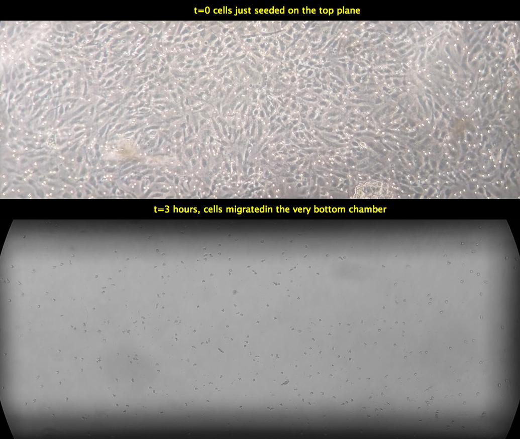

Although these are the representative images, I did see more migration the case of control devices. I had 2 controls and 2 blocking.
In the control device, I have the cells on the top layer, then as I scroll through the 600 um collagen gel, I don’t see any neutrophils in the intermediate planes, but all the cells that have migrated are all in the bottom channel.
However, in the case of blocking CD29 on neutrophils, I could see many cells in the intermediate planes, and a decent number in the very bottom plane as well. It implies that neutrophils were able to beat the inhibitory effects of beta-1 blocking Ab, but it took them time and reduced the speed of their migration through the collagen layer.
Summarily, I quantified the cells in all the 4 devices: I counted cells in the bottommost planes 3 hours post seeding, and divided with total number of cells in the topmost compartment when cells were just seeded, and took the ratio of former to latter and reported it as ‘percent migration’, as shown below.

This week, I am going to repeat the same experiment to ensure more statistical robustness. Stay tuned!
UPDATES: October 31, 2017
I pooled more data for the above experiments from earlier dates and last few weeks. I will update the statistics later on the blog towards the end.
As I mentioned in the paragraphs above, I am using collagen gel in both the apical and basal chambers. The gel on the top is to promote cell adhesion and providing a compliant substrate for cell culture, while the basal gel is to mimic ECM space. Technically speaking, I don’t need the bottom gel since I am not studying interstitial or 3D migration. Having bottom gel makes my job difficult in quantifying and imaging the neutrophils in the bottom chamber. Cells scattered in all the focal planes diffuse the signal and are also prone for miscalculating. So I decided to study how the migration counts differ if I reduce the complexity by having only the top gel and not the bottom gel.
I am uploading again 2 representative videos of control vs block case.
CONTROL CASE
BLOCK CASE
As we could see, very few cells transmigrated in the bottom chamber in the beta-1 block case compared to control case. No cells are observed in the intermediate planes because there is no gel in between.
Finally, I compared the data from top+bottom gel with top gel only case for control vs block conditions.
Statistics are self explanatory.
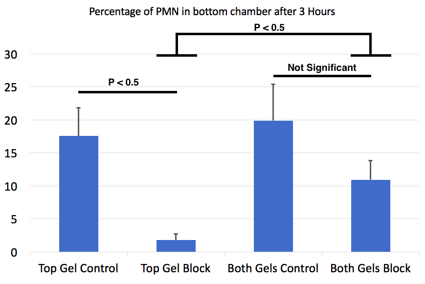
As we can see in the top gel case, the control and block differ significantly, but this difference between control and block is absent in the case of both-gel case. Fortunately, removing bottom gel and blocking neutrophils prevents their migration significantly as seen by the difference in column 2 and column 4. Few speculations to explain these differences:
- The absence of bottom gel makes neutrophils harder to fall from the trench in the bottom chamber. the glass coverslip is 730 microns away from the trench when the bottom gel is absent, but this distance is only 130 microns in both-gel case. Probably the bottom gel provides an easier access to chemotax for neutrophils and the falling down is conducive. Maybe, just a thought?!
- Absence of bottom gel exposed the basal fMLP directly below the membrane, making the gradient very shallow. Again in bottom gel case, fMLP is spread over 6+1=700 microns below the cells, whereas this gradient is only 100 microns wider. Lesser gradient = lesser motivations for neutrophils to chemotax = lesser count of cells in the bottom chamber. Speculation, but stronger than point 1.
As always, stay tuned for some more microscopy based evidence of cell migration!
This is heroic, using an antibody in this context is likely to lower your active concentration through the collagen acting like a big sponge.
Have you considered parallel efforts using siRNA or antisense to knock down the mRNA and prevent expression that way?
Antisense on human neutrophils freshly isolated from blood? Is it possible?