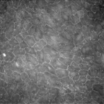
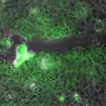
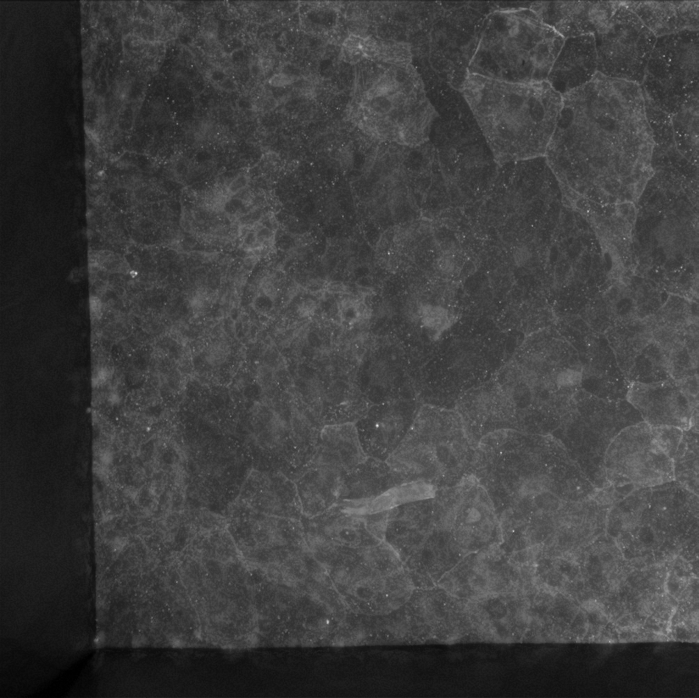
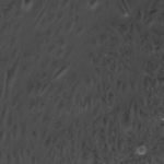
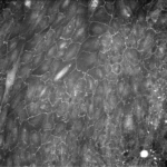
RPE ZO-1 expression observed on MgF2 and tissue culture plastic
Here are some more images from my ongoing cell culture project.
My previous work and protocols: ZO-1 staining of confluent RPE cells on various MgF2 substrates
I added twice the amount of primary antibody on these devices that I usually do (1:100 dilution, instead of 1:200).

High Porosity MgF2
TCP
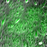
MgF2 Coverglass