Dead End Filtration of Gold Nanoparticles Update
Two weeks ago (as of the time of writing this post), I wrote a post on a dead end filtration experiment that I ran in order to demonstrate particle capture in pores, modeling a simple, purified exosome capture system that we could use for imaging or analysis. However, when I ran that experiment, I done goofed and suspended the nanoparticles in PBS, which causes them to aggregate. This messed up the results and caused the formation of large masses of the nanoparticles on the top surface of the membranes. A few of the nanoparticles were in pores, but the pores were largely unoccupied.
(Note: The wafer used in these experiments was 2226, which was 50 nm NPN with 50 nm pores.)
Yesterday, I repeated the experiment and this time I suspended the gold in nanopure water. I used the same ladder (30 nm, 40 nm, 50 nm, and 60 nm) and the same membranes (50.7 nm average pore diameter, 13% porosity) and the same speed in the centrifuge (5 min at 2500 rpm). The resulting data represented more of what we expected to see from the initial experiment. For the membrane with the 30 nm gold, as we can see in Fig. 1, almost no particles were retained and they all passed completely through the membrane.

Figure 1: SepCon membrane with 30 nm gold particles. The membrane is clean because the gold passed through. Only a few remained in the pores.
The 40 nm gold started to approach the size of the pores and we could see that many of the pores were occupied by nanoparticles as we can see in Fig. 2. Sometimes, there were two nanoparticles per pore, but most of the time it was only one.
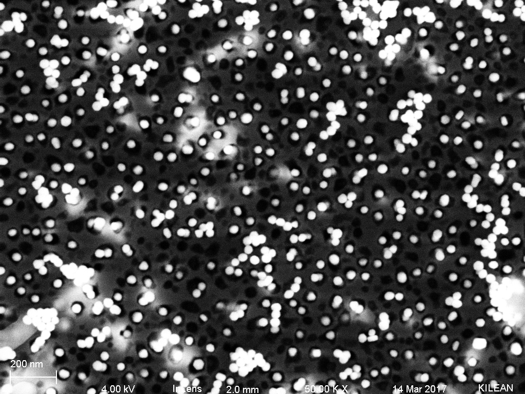
Figure 2: SepCon membrane with 40 nm gold nanoparticles. Note how the majority of the pores are occupied. Sometimes, there are multiple particles per pore.
The 50 nm gold was approaching the size of the pores and as we can see in Fig 3., almost all of the pores are occupied, typically with 1 nanoparticle per pore.
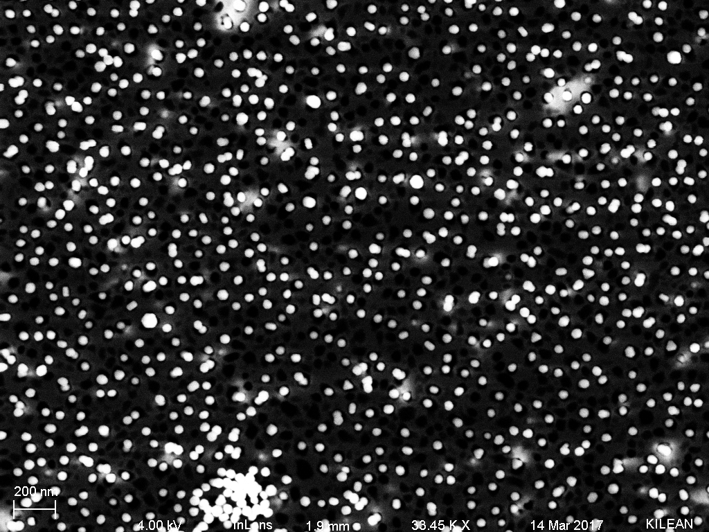
Figure 3: SepCon with 50 nm gold nanoparticles. Note that pore to particle ratio is almost 1:1.
Finally, the 60 nm gold particles occupied some of the larger pores, but the density was noticeably lower than the 40 and 50 nm gold. We can see this in Fig. 4.

Figure 4: SepCon with 60 nm gold nanoparticles. The pore occupation is significantly lower, indicating rejection unless the pores were the right diameter.Note how the larger pores all appear to be occupied.
Given these results, it should be fairly straightforward to capture purified exosomes on a SepCon style membrane in dead end filtration. The exosomes can then be imaged or further analyzed however is seen fit.
Nicely done. Agreed. For purified exosomes, SepCons should do the trick. Time to get some purified exosomes …
At some point we need to do these experiments with classic pores (slightly tapered) to illustrate the bowl with a whole catch concept.
Can you update this with the concentration of particles used? It is critical to get that right and they change as we change the size. If it is in your prior post please provide a link to that post. Actually it is always good practice to link to prior posts that are part of the thread.
Should these NPs be measured for their size distributions? I could imagine that presenting the size distributions of the NPs side-by-side with the pore distributions would help make the point. Interesting results though. Did the researcher in urology give you some ideas about the usefulness of capturing purified exosomes on these membranes?
Please put the wafer number in this post.