Hand-cast Protein Gels: New Technique and Fresh Chemicals
In April of last year, I spent a considerable amount of time trying to visualize protein separations using SDS-PAGE (Sodium Dodecyl Sulfate-PolyAcrylamide Gel Electrophoresis) gels, and ultimately was able to get some data:

I was trying to show that 15 nm pnc-Si could be used to separate BSA from cytochrome c, and while at first it looks like I did exactly that (and indeed, that’s how the figure was labeled), when I looked closer at the data I realized that what I was actually showing was a separation of dimerized BSA from monomeric BSA, and the cytochrome c had run off the gel completely:

These images were taken using a pre-cast gel that I purchased from the Biological Supply Center in the medical center. I had tried to make my own hand-cast gels using the buffers and reagents that we had lying around and using a protocol that Jess had written up, but I could never get my gels to polymerize. I bought some new TEMED and messed with things a little bit, but at that time I had other projects happening and I was hesitant to update everything.
For the sake of Jess and Paul Black’s paper that we’re trying to rescue from purgatory, I’ll need to be making a fair number of protein gels, and I returned to the problem. This time I used a different protocol, one from Cold Spring Harbor’s website, and made everything (concentrated resolving & stacking buffer, 10% SDS, sample buffer, APS, bromophenol blue, glycerol, stain, destain, etc.) fresh, and purchased new Acrylamide solution. From mixing the resolving gel to imaging the destained gel took me about 5.5 hours. Because this was just a test of the chemicals and technique, I didn’t do a separation with my protein mix. The protein mix I used was as close to the one Jess used as possible – 0.5 mg/mL each of carbonic anhydrase (37 kD), ovalbumin (45 kD), and albumin (67 kD). I also included 0.26 mg/mL of phosphorylase b (195 kD), because that was all that was left. I did not include  -galactosidase (495 kD) because we don’t seem to have any left. The proteins were in 10 mM KCl. I stained the 10% gel with coomassie blue:

UPDATE: I left the gel in destain overnight, which gave me a better image:
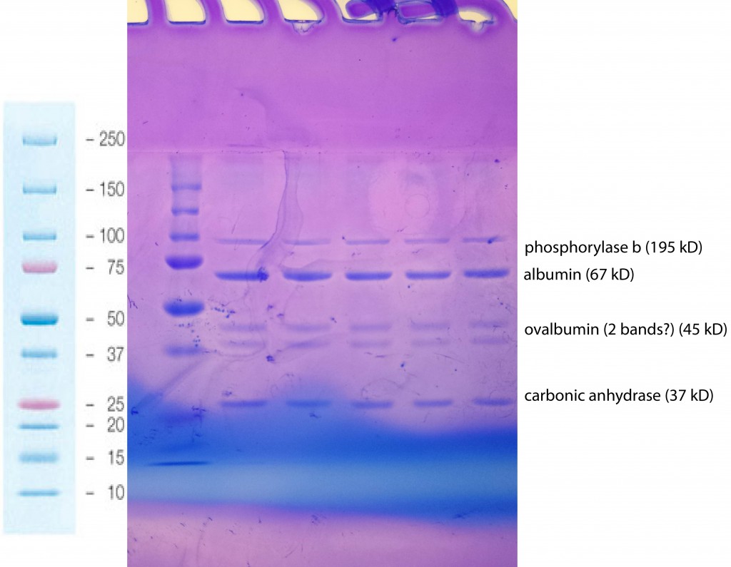
There’s some weirdness around ovalbumin (I think there’s two bands?), and it’s obvious now that I should have let the gel run for another forty minutes or more. I believe I ran the gel for 120 min, but I can’t be sure, because twice during the run the buffer in the inner compartment of the cell either leaked or boiled off, breaking the circuit and stopping electrophoresis. I don’t know exactly how long each of these breaks lasted for. Next time I will be more careful in monitoring the cell.
Recipe (including recipes for buffers, staining solutions, etc., as well as an overview of the technique)
Website Part 1 (SDS-PAGE of Proteins)
Website Part 2 (Staining Proteins in Gels with Coomassie Blue)
Note that Cold Spring Harbor’s online protocols are only available when you are logged into their server. The Windows machine in the alcove is apparently signed in, but I wasn’t able to bring it up on my personal mac, even after signing in to the library website.
If you run into trouble with the Bio-rad mini-protean device itself, consult the manual.
How long did you run the gel? Another 40 minutes sounds like a very long run. If memory serves, the typical time to run a gel is 30-40 minutes.
It also looks like you should have destained longer. The gel background is too dark.
I believe I ran the gel for 120 min, but I can’t be sure, because twice during the run the buffer in the inner compartment of the cell either leaked or boiled off to below the edge of the wells, breaking the circuit and stopping electrophoresis. I don’t know exactly how long each of these breaks lasted for. Next time I will be more careful in monitoring the cell.
I left the gel in destain overnight and have updated the post with the (better) image I got this afternoon.