PDMS cell channels
Michael and I have been working to create cell channels of varying widths and heights to test out a concept used in a paper we recently read as a group. This work claims to see cell cords in 12 hours, so no media renewal is necessary.
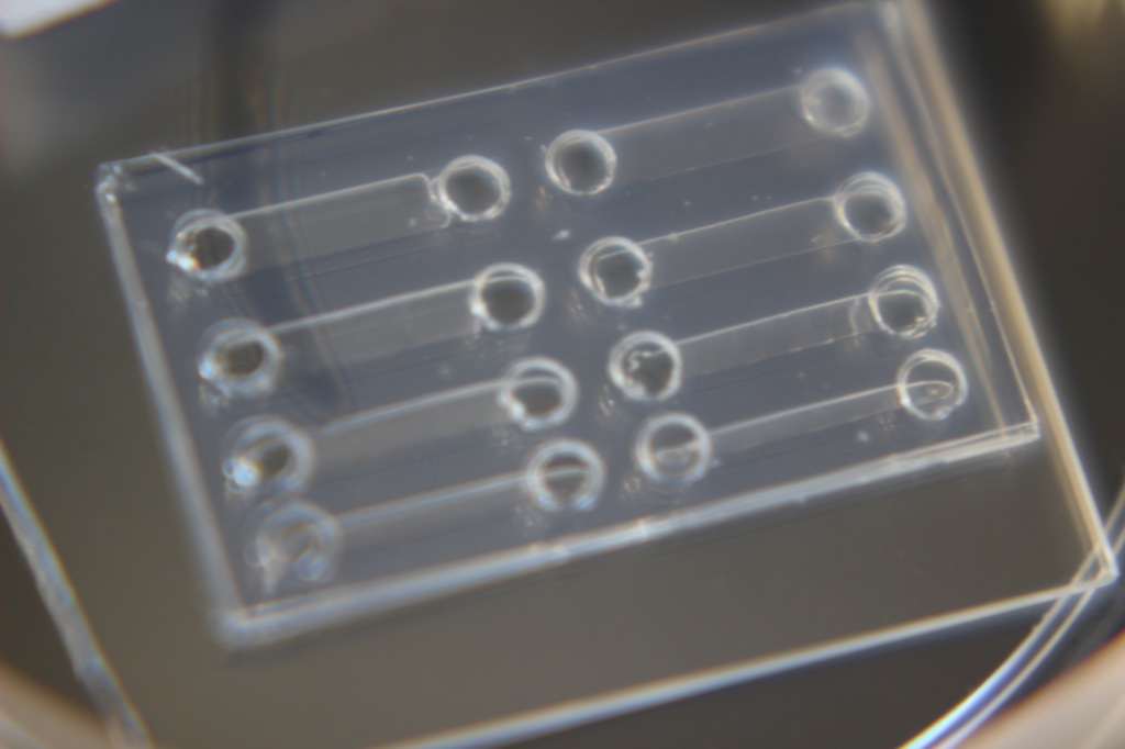
The channels were made by layering and bonding PDMS onto a glass slide. The first layer containing the channel cutouts can be made with either 100 um or 300 um PDMS, and then 2 layers of 300 um PDMS were placed on top, with loading holes at the beginning and end of each channel. The thicknesses we chose to test are 500 um, 1000 um, 1500 um and 2000 um. The length of each channel is 10 mm.
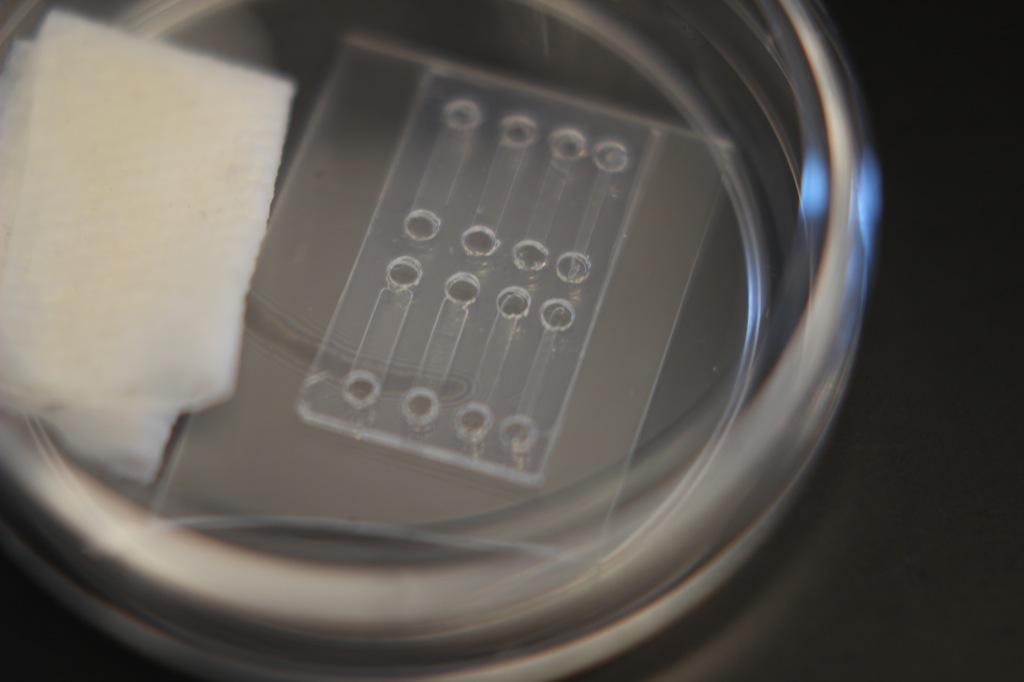
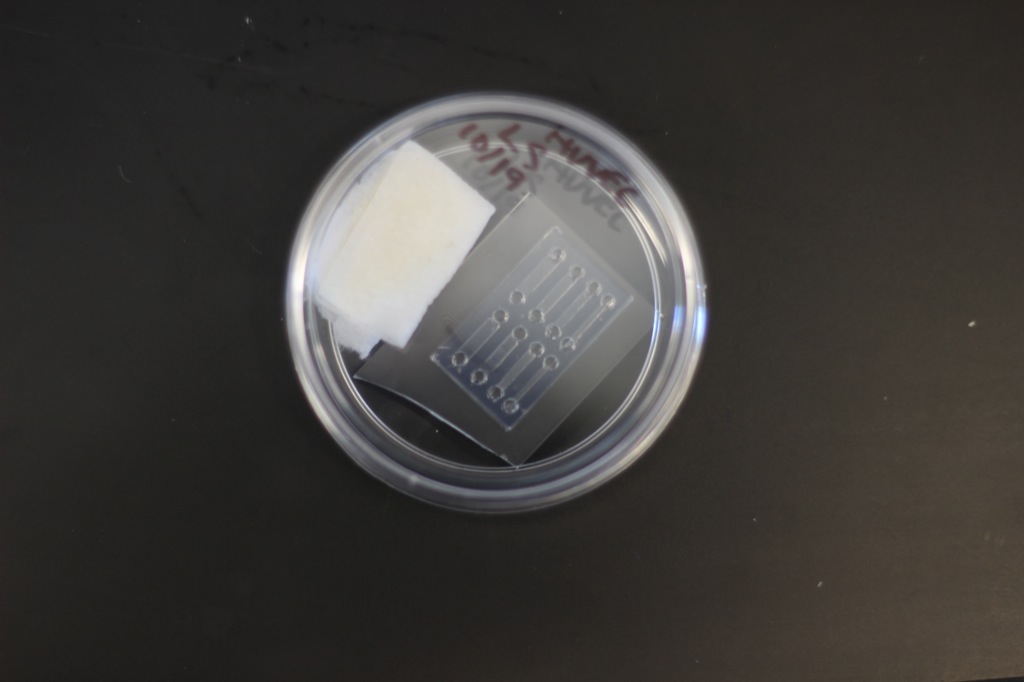
The method for mixing and loading cells was taken from the paper. We mixed equal volumes of 3 mg/mL type 1 rat tail collagen and HUVECs in medium 200. This brings the final collagen concentration to 1.5 mg/mL and the cell density becomes half of what it was before mixing. This is kept on ice and pipetted into the channels, and then allowed to gel in the incubator. We moved the slide to a petri dish and added a saturated kim wipe to reduce drying/evaporation.
- Paper used 5 mg/mL for a final concentration of 2.5 mg/mL, but other literature suggests final concentration of 1-3 mg/mL is optimal.
We have not seen tubes/cords form yet. This is what the channels look like upon seeding:
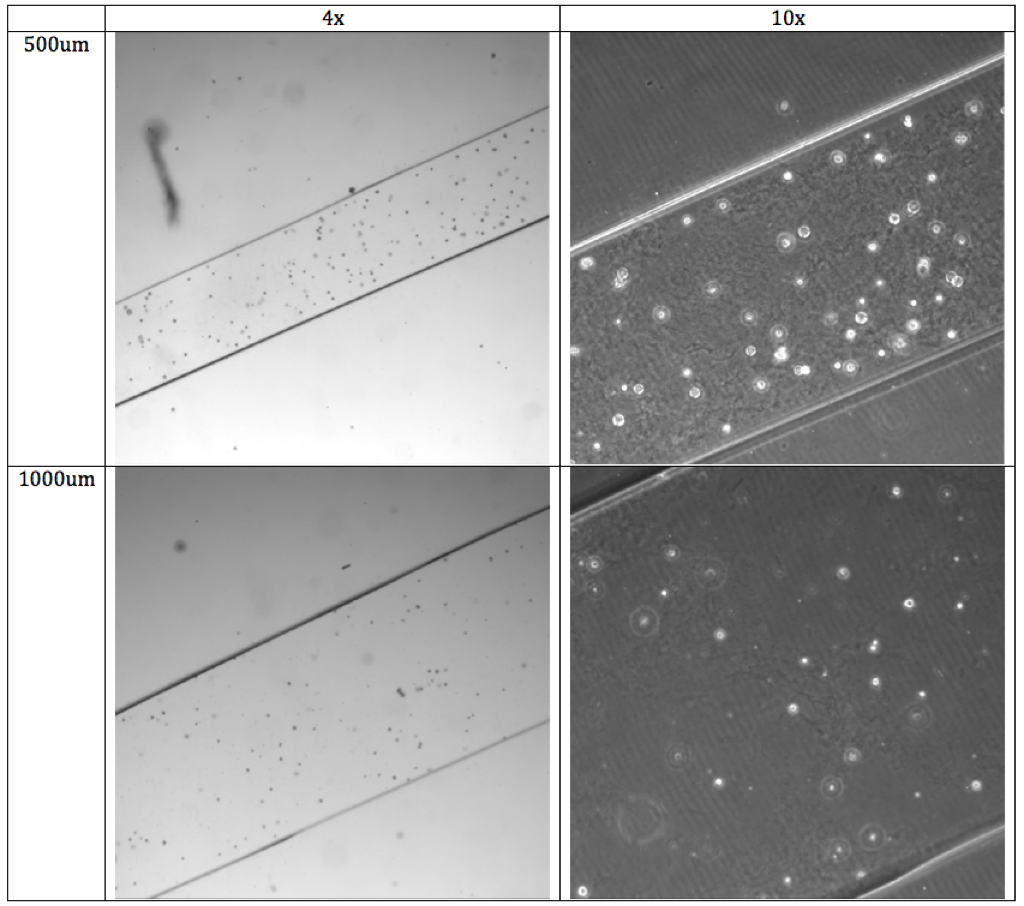
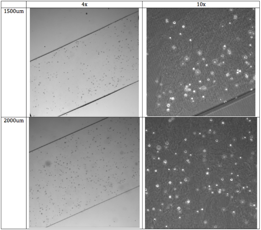
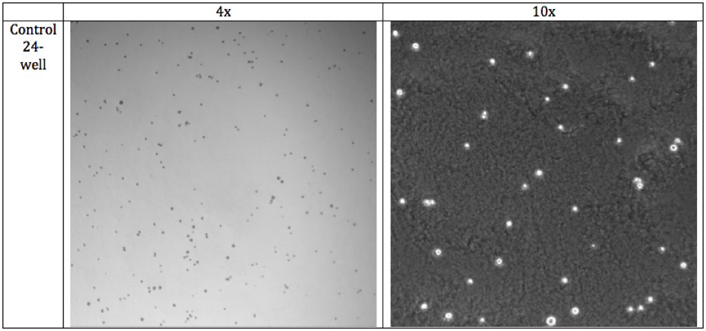
The cells seem to stay rounded up the entire time. I am wondering if this has to do with pH issues, as usually I have diluted the pure collagen with DI water, sodium bicarb and 1X MEM.