Low density bEnd cells have no vacuoles on pnc-Si
In all of my experiments involving vacuoles, I’ve seeded at 50000 cells/cm2 which gets the cells to confluence after ~ 1 day of growth. I’ve always seen vacuoles with these parameters. For this experiment, I wanted to see what the cells would look like after 1 day but at a much lower seeding density. The question is : is vacuole formation a function of polarized endothelial cells or will single cells express vacuoles? In vivo, endothelial cells have 2 distinct ‘sides’ – the side facing the bloodstream(luminal) and the side facing the ECM/other vessel wall cell types (abluminal). This 2-sided phenotype (also a characteristic of epithelium) is called ‘polarization’. For polarization to occur in vitro, the cells must be on a permeable substrate, although I don’t think that most cells will polarize by themselves on transwells without stimulation by an exogenous signal.
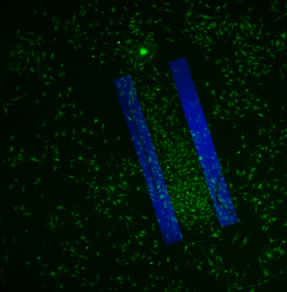
Anyway, here I seeded P15 bEnd3 cells @ 10000 cells/cm2, so ~1/5 that of normal. I allowed them to grow for 1 day and then stained with Live/Dead. I imaged 2 different samples which looked essentially identical, so I’m only including one here.
This 20X image shows a fairly low density of cells that are fairly spread out and almost spindly. These cells are clearly not confluent yet and they’re still crawling around. There are no vacuoles in any of the cells over the blue membrane. Unfortunately, I’m not sure what the timecourse of vacuoles/confluence/polarization is here. I find it hard to believe that high density bEnd3 after 1 day (my ‘typical’ experiments) polarize and then express vacuoles (it seems too fast). Therefore, do the cells reach confluence, experience some contact inhibition and then express vacuoles as they polarize, or do they express vacuoles long before they polarize, or are they even polarized at all? I guess this experiment has opened up a few more questions than it’s answered.

However, this lower magnification (4X) image is pretty interesting. Notice the extremely high density of cells around, on and in between the membranes compared to the rest of the sample. Unless this is a seeding artifact, this is pretty strong evidence that cells are being recruited toward the free-standing membranes. I need to get fluorescent time-lapse movies working!