Vacuoles don't co-localize with nuclei
I recently started up another BBB co-cultures and I had a couple of leftover samples. I decided to use one for a ‘just-to-be-sure’ experiment since I realized that I never did a calcein-Hoechst 33342 co-stain. That is, I never used a nucleus-specific dye with calcein to confirm that the black holes (vacuoles) in the green channel were not nuclei. In phase contrast images, it seemed like the vacuoles were unique structures and I had no trouble distinguishing them from the nuclei. However, this proves it in color.
I seeded P10 bEnd3 cells at 50000 cells/cm2 (normal density) on 2×2 square membranes from SC604 and allowed them to grow for 1 day. I then made the normal Live/Dead solution in media but supplemented it to 1uM with Hoechst 33342 (a blue, nucleus-specific dye). I incubated at 37C for 30 minutes, rinsed in PBS and then imaged on the Zeiss.
The following is a 3 color (calcein-green/Ethidium-red/Hoechst33342-blue) overlay. In no instance does a blue nucleus co-localize with an unstained vacuole in the green channel. The holes definitely aren’t vacuoles.

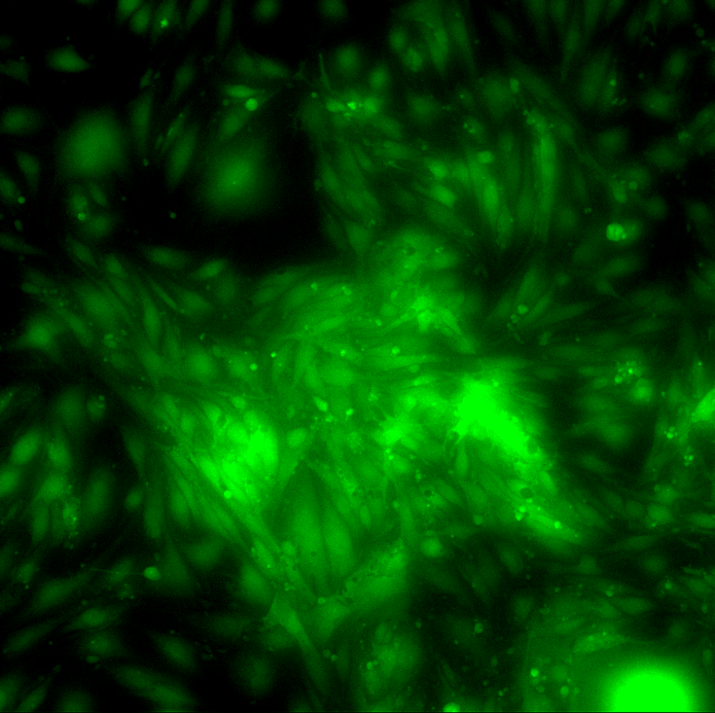
This is more like what we’re used to looking at, the green channel only, for comparison.
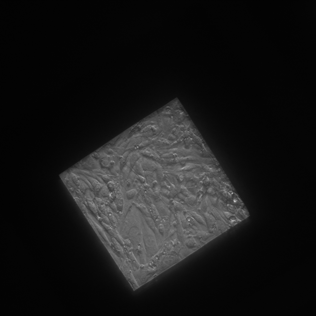
In case you couldn’t figure out where the membrane was, here’s the Phase:
Extra thought (March 31):
The image below is just the nuclei for the same region of interest as above. I believe that you can tell where the membrane is just by the ‘density’ of nuclei per unit area. I think if we want to quantify cell density over different areas of pnc-Si, counting nuclei will probably be easier with the matlab cell counter than calcein-stained cells.

I love the nuclei only picture. Yes its a better way of doing counting especially in crowded cultures.