Track Etched SEM
Because of the disagreement between the experimental and simulation comparison in this post, I decided to take a look at the Sterlitech membranes using SEM. Barrett gave me a hand with all the images, and we tried to look at the edge for thickness and the pores for density/overlapping pores.
In this first image, the membrane is folded so that you can see the edge straight on. The methods used to cut the membranes may mean that the edges may not represent the thickness, but it was an easy first thing to look at.
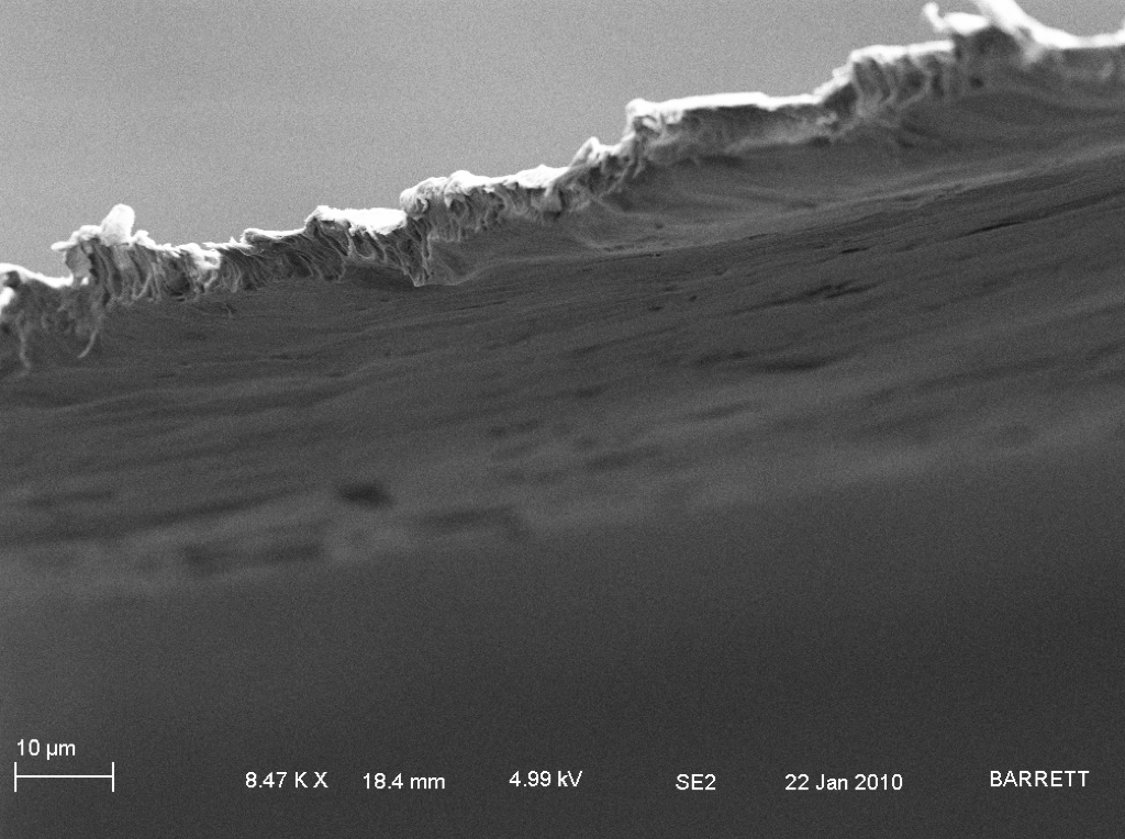
By a rough px/nm conversion based on the scale bar, the edges here are between 3.2 and 5.7 um. The company says the membranes are 6um +- 10%. Thickness plays a big role in the simulation, so if it is thinner than they say, my results would agree better with the theory.

I still need to try some pore processing, but pores may be slightly bigger than 30nm as the company sells them. However there don’t seem to be too many overlaps and the pore density is about what they say it is.
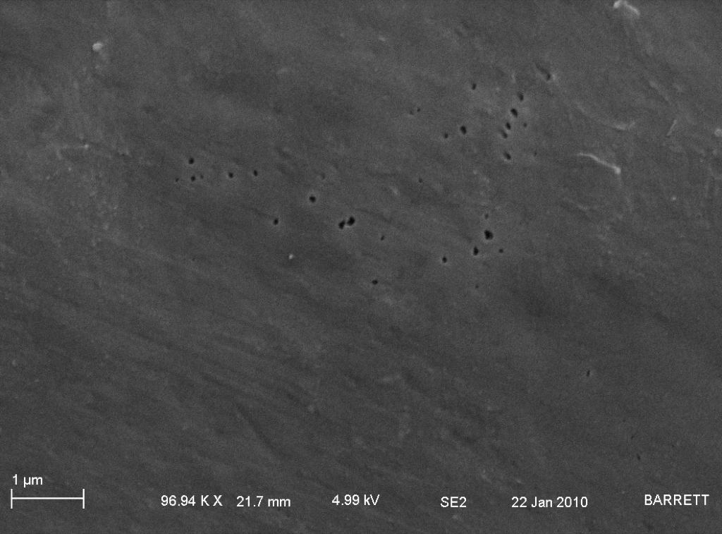
The pores appeared to be in patches especially near the edge of the membrane. I’m not sure why this is so, but maybe they make each membrane disk separately or there’s issues with etching at the edges.