Electrophoretic Cell Update
We were worried that Friday’s experiments were giving us a false positive results. If the electrophoresis breaks the membrane, then we’d expect to see the rhodamine rush through just like it was in our experiments. It’s too hard to view the membranes afterward because they tend to break during removal. I designed some simple tests to verify if they were in fact intact.
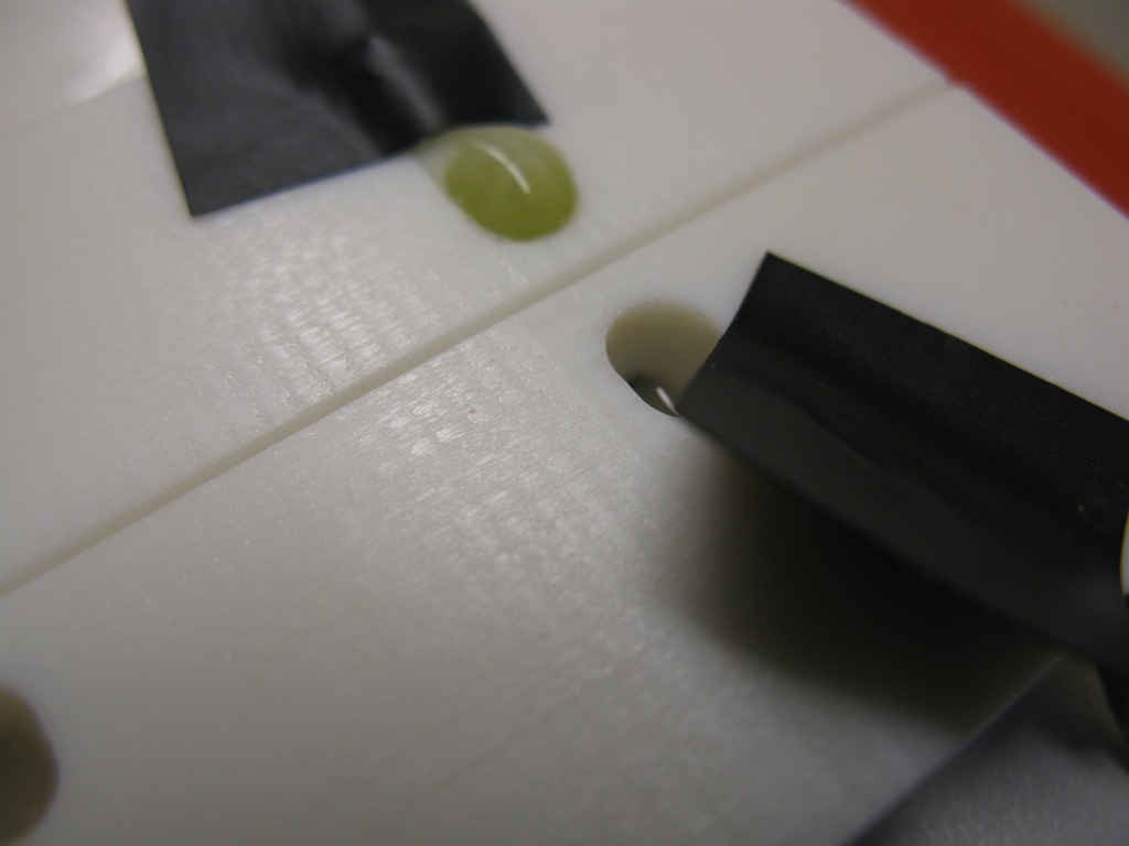
I set up both experiments with .5mM Rhodamine and PBS. For the first test I ran the setup using 20V (8mA) for 5min. The filtrate well reached a faint pinkish color, while the retentate remained red. In the case of broken membranes, equilibration is practically immediate. If the filtrate remained only pinkish, then I would know that the membranes are still intact. In the second experiment I ran the system at 20V(8mA) for 1min then removed the slightly pinkish filtrate and replaced it with clean buffer. If the membrane is broken, the fluid would pour into the filtrate after it had been removed and quickly reach equilibrium with the clean buffer.
Both membranes passed the tests. Here is a picture from the second. The filtrate looks a little pinkish due to some staining of the stir cell, but it never appeared any darker over ~10min.

Retentate left, filtrate right.
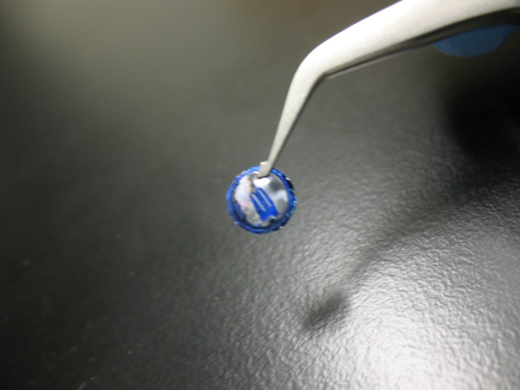
After these tests I started to fiddle with the system (typical, I know). On the first experiment setup, I tried to reverse the polarity and send the rhodamine back to the retentate well. Nothing was happening and the system wasn’t reading any current. Worried that my wires got detached, I spun it around and tried to run it in the usual direction. Again nothing happened. I tried to run the second experiment again in the usual direction and nothing happened here. Frustrated, I removed the membrane and meant to put new ones in the stir cell. As I pulled out the membrane I was surprised to see that it was glowing pink! Rhodamine must have stuck to both of them like crazy and plugged all the pores. I’m not sure if it’s because of electrical interactions or cake formation. Here’s a pic:

I rinsed the membrane in PBS and then pentane. While it partially broke, the remaining fragments of the suspended membrane were still bright pink. I then imaged the remaining fragments with the fluorescent scope:

The membrane appears to be brighter than the surrounding pnc-Si. Maybe it’s an optical artifact, but I guess if the rhodamine attaches internally and to both sides it could be this bright.
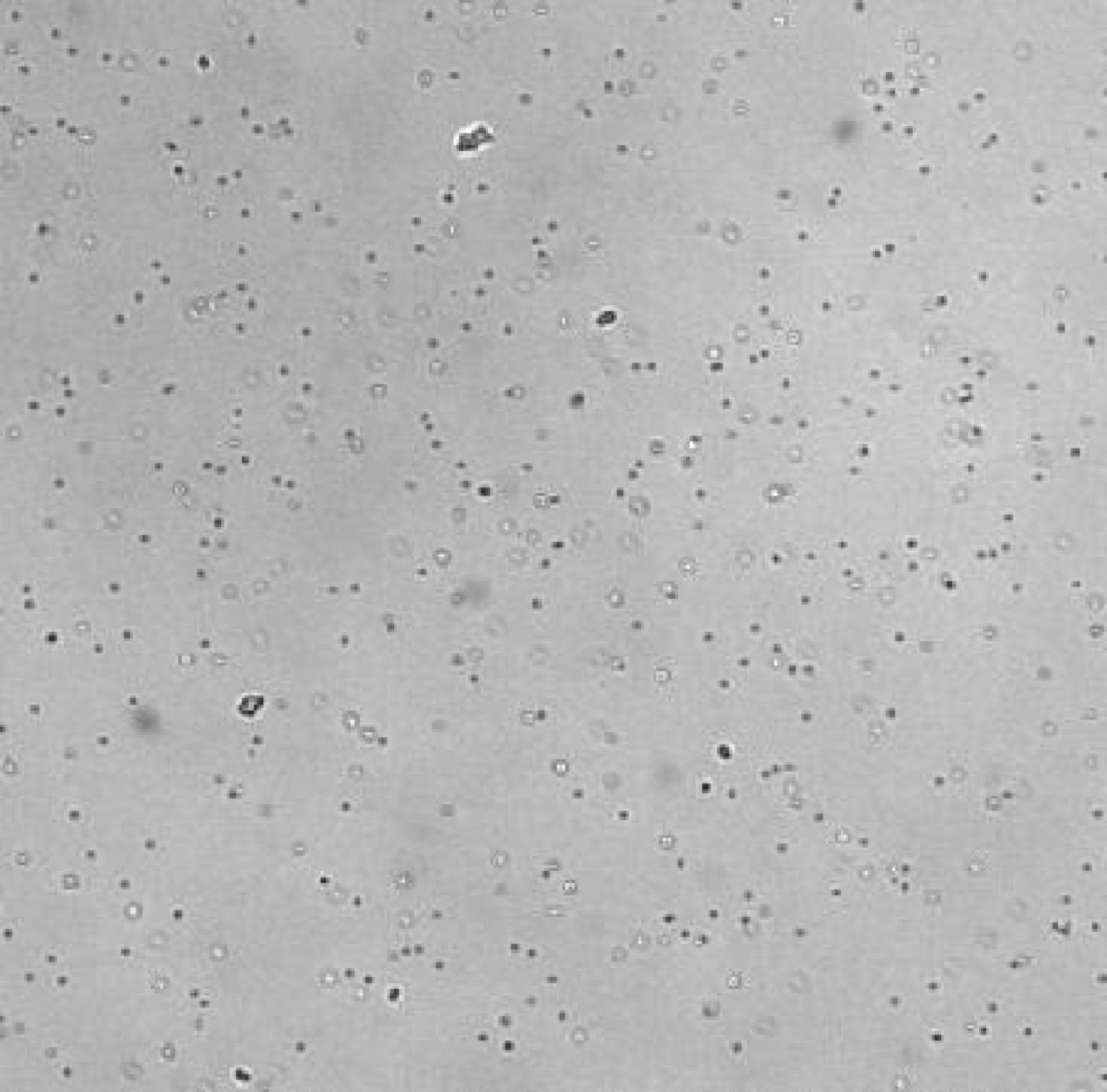
I also performed a separate but similar experiment at Tom’s suggestion. I set up two tests, one with a broken membrane and one intact (but with 3 pinholes). The retentate wells held 1:100 1um yellow/green beads in PBS and the filtrate wells held PBS. I set up the field on the broken membrane first, and after 5min sampled the filtrate and observed it using the scope. I could see many beads in the sample:
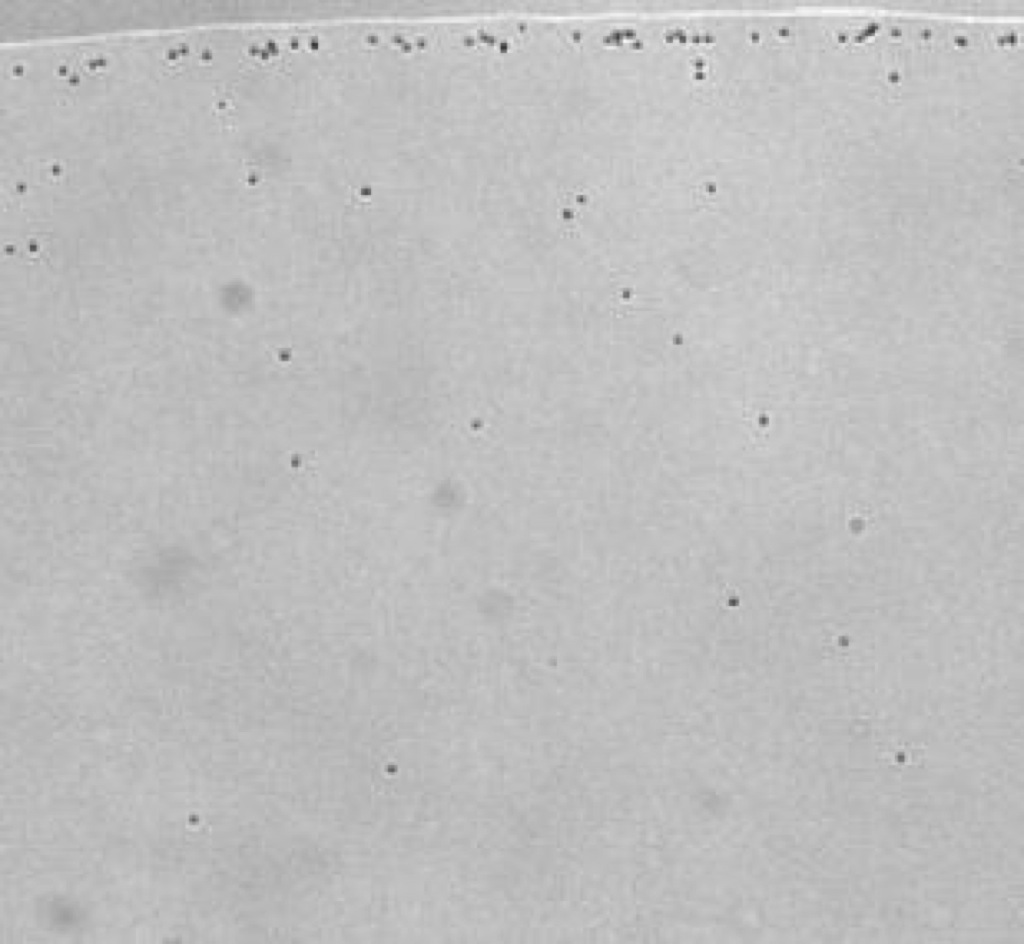
I did the same with the pinholed membrane. There were few beads in the sample and most were clustered at the edge of the drop:
I removed both membranes to view using the scope, but I was surprised to see that the pinholed membrane appears to have discolored:
You can see the edges of the o-ring and some color still surrounding the slits. I’m not certain if this is our usual discoloration or some sort of electroplating phenomenum. I checked the membrane using the scope. Here’s the front side (which is in contact with the retentate):
 DIC
DIC
 FITC
FITC
Here’s a view of the surface away from the suspended membrane:

You can see the individual beads in this image and some sort of front.
Then I looked at the backside (well side). So this is the pinhole membrane and we don’t expect that many 1um beads are making it through.

One other strange thing that happened was that when I let the pinholed membrane continue to separate beyond the 5min sample, the fluid all started to go toward the retentate well. It started poring over the top of the stir cell while the filtrate got lower and lower.
Retentate left, filtrate right.
Changing polarity on bead system:
So Dave’s question was would reverse polarity cause the same sort of discoloration? I tried this with everything set up the same except the electrodes reversed. There was no discoloration of this membrane after running for 10min in this manner. The retentate beads however lost their color and the filtrate well swelled this time:

Retentate left, filtrate right.
The beads however maintain their fluorescence and don’t appear to be super clumped under the scope.
Do you observe similar behavior (discoloration on the front) when you reverse the polarity. Is one side more basic than the other? Since you’re introducing a potential across the membrane, ions will separate. Could the increase in fluid level be from osmosis?
I’ll try reversing the polarity. The only differences in the solution is that one is PBS and one is PBS + a couple uL of beads.
Now I’ve considered osmosis since you’ve got all these beads on one side, but since the ions can easily move across the membrane they will balance out that difference. But maybe since we’re actively drawing the ions that might change things. Electro-osmosis is another possibility. This is where you line up charges on the wall of a tiny channel and get transport of a polar liquid by plug flow I think. Maybe we’ve achieved this even though we didn’t think it was possible in our hole-like geometry?