Attempts at Co-culture
Attached is a series of images we took of two populations of neutrophils above and below the membrane.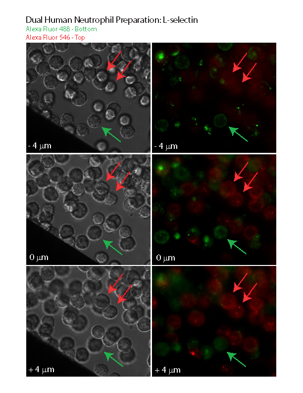
I’m including by methodology for those who are interested:
One population was pre-labeled with AF488 (green) antibody against L-selectin, the other with AF546 (red). The green population was suspended in a low protein buffer (~1% serum) and a 20 uL drop was placed on the smooth surface of the membrane. I let these cells settle for about 15 minutes. I have previously experimented with different protein concentrations and found the membrane acts like glass – if my buffer has >2% serum, then neutrophils will not easily adhere and just rest on the surface. Since I needed these cells to lightly stick to the membrane, I put in a low protein. No protein typically results in the cells activating and spreading on the membrane (or glass). Next, I placed two small dabs of vacuum grease (w/ 50 um silica beads) on a clean coverslip. I pipetted off most of the green cell solution and flipped the membrane onto the coverslip so that the flat side was now facing down (suspended by the beads in the vacuum grease). The green cells are now hanging from the membrane. I then pippetted red labeled cells with the same 1% serum buffer into the well (about 2 uL).
I took a series of images every 1 micron using our 100x objective on the Zeiss microscope. I selected images that I believe to be 4 microns below, at the membrane and 4 microns above. In the DIC (b/w) images, you can see one population of cells that are in focus, while about half appear out of focus. At the membrane, they both appear to be slightly out of focus. But it is clear that some of the cells are out of focus because they are above and some are out of focus because they are below. Then The last DIC image shows the inverse of the first image with the other (red) population in focus.
Lastly – a big thanks to Henry for donating his blood and performing the neutrophil isolation.
I assume that 1% albumin is 10 mg/mL – I guess this is “low protein” relative to pure serum. It’s interesting that you see any difference in solutions with this much protein. Do you think it has to do with the surface or the cell?
I meant to say 1% serum. I edited the blog to reflect this.
And thats a volume % not mass. It should be << 10 mg/ml.