HUVEC Mono(?)layer Na-F Permeability
I had some old HUVEC (P8) on pnc-Si transwells and PET Costar transwells that I didn’t know what to do with. So I tried a permeability study to see if there were any unexpected roadblocks in the protocol.
I seeded HUVEC @ 50,000 cells/cm2 on PET and pnc-Si and allowed them to grow for 6 days. I then washed the inserts with HBSS+ (+Mg, +Ca, +10mM HEPES) once, then incubated the inserts in this solution for 15 minutes @ 37C. It’s important to used HBSS with Ca and Mg because intercellular junction proteins are calcium dependent. I then moved the inserts to new wells with 1mL of HBSS+ and took the 24-well plate into the Tecan (heated to 37C) for a measurement without fluorescein. I then added 200uL of 0.00075% Na-F and took measurements every 15 minutes with a manual gain = 86. I actually used the wrong plate definition for the 15 minute time point, so the data starts after 30 minutes of transport. This protocol involves simply transferring the plate from the incubator to the heated Tecan (without removing the plate, obviously) for a measurement and then putting the plate back into the incubator. No sampling required!
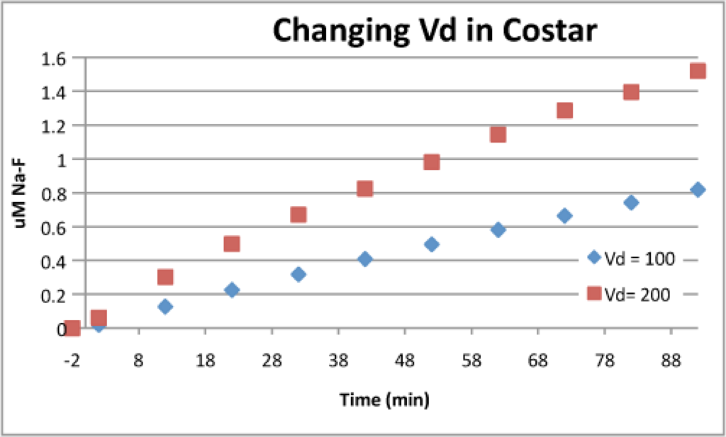
The following graphs show that HUVEC slowed Na-F transport through the PET and pnc-Si transwells. After 90 minutes, Na-F transport with HUVEC was ~ 66% of the acellular transport in PET and ~ 20% of the acellular transport in pnc-Si. This means that pnc-Si is more sensitive to cell monolayers than transwells. For some reason, Na-F transport was fast for the 1st 30 minutes but slowed down considerably after that in the HUVEC sample. I should also point out that the acellular data for pnc-Si is extrapolated. As of now, I only have pnc-Si transport data with donor volumes of 100uL. The cellular studies were done with a 200uL donor volume. I’ve found that the transport increases linearly with increases in Vd (see 3rd graph below), so I multiplied the acellular data by 1.5 (to be conservative). Therefore, this data only gives us a qualitative idea of transport through monolayers with our set-up. However, it’s important to note that fluorescein transport through transwells is plenty fast enough to pick up with the Tecan protocol I designed. We’re definitely ready to take cell monolayer permeability data with the Tecan.

Transport increases ~ linearly with donor volume (Vd):
Immediately after the transport experiment, I stained the cells with CMFDA, a viable cell dye and then fixed them for observation. Below are 2 images of HUVEC on different areas of the PET transwell.


The cells don’t look all that great, and there is certainly not a confluent monolayer. It’s interesting that there was a fairly significant decrease in transport even though the PET membrane was not covered by a confluent HUVEC monolayer. It’s possible that this protocol stressed the cells too much (they had been in serum-free media for about 2 hours by the time I stained them), but it’s also possible that the passage number was too high for confluency. It’s promising that cells can be stained after performing a transport experiment – this means one transwell device could be used for 2 different characterization experiments.