ZO-1 Staining on ARPE-19 Cells grown on MgF2 nanomembranes
The following images are from ARPE-19 Cells cultured for 3 Weeks over here in Nottingham (thanks for help from Emilia Moradi, Victoria Ciampani, and Kevin Webb).
MgF2 nanomembrane chips were positioned into a transwell insert and sealed using PDMS, a biocompatible polymer, creating a two-compartment transwell. After an ethanol sterilization, ARPE-19 cells were seeded in a 12 well plate (100,000 cells/well) on MgF2 nanomembrane inserts and polyester transwells (Transwell® COSTAR 3462, Corning Inc.), having previously incubated the substrates in media (DMEM:F12,penicillin (100 U/mL), streptomycin (0.1 mg/mL), amphotericin (0.25 μg/mL) and Fetal Bovine Serum (FBS, 10% v/v,)) for 3 hours. Cells were grown over 3 weeks in an incubator (37 oC, 5% CO2) to confluence on these substrates, exchanging the media every other day.
Confluent cell monolayers were washed with PBS and fixed in 4% paraformaldehyde for approximately 10 min at room temperature. Cells were then washed 3 times with PBS and permeabilised by incubating with Triton X-100 (0.1% v/v in PBS) for approximately 10 min. Cells were then washed with PBS, followed by the application of 1% BSA/PBS for approximately 1 hour. Thereafter, BSA/PBS solution was aspirated and replaced with mouse, anti-human ZO-1 (primary) antibody, diluted in 1% BSA/PBS. Cell samples were incubated with the primary antibody overnight in the fridge. The primary antibody solution was then removed and cells washed with PBS (3 times). FITC-labelled goat, anti-mouse (secondary) antibody, diluted according to manufacturer’s instructions in 1% BSA/PBS was then applied to the cells for 1 hour. The secondary antibody solution was then aspirated and cells washed with PBS extensively. The Transwell® filter membrane was excised and mounted on glass slides (using DAPI-containing, ProLong® Gold antifade/mounting medium). for confocal imaging, which was performed using a Leica TCS SP2 system mounted on a Leica DMIRE2 inverted microscope. Image stacks were reconstructed using ImageJ.
Below are some representative gifs that go through the confocal z-slicing as they were acquired. The ‘scanning’ behavior shows that the MgF2 substrate is tilted. There is some jitter and drift over the 30-45 minutes these stacks were acquired.


Shoving the images through a stabilization scheme produces: http://www.cs.cmu.edu/~kangli/code/Image_Stabilizer.html
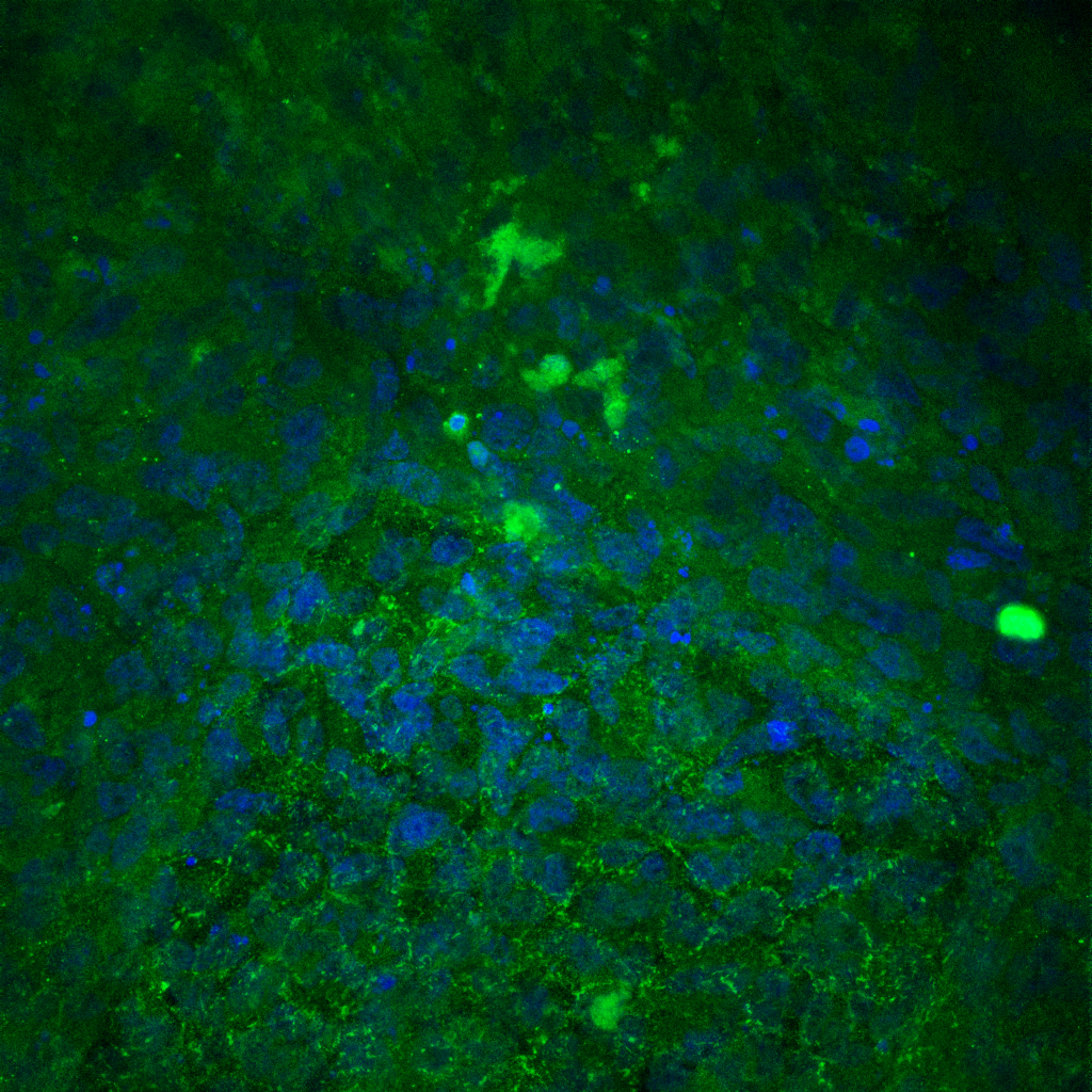
Here it is easier to see some TJ expression away from the permeable region in the lower left corner, as well as the upmost point on the MgF2 square.