DAPI & Phalloidin Update – Thicker SiO2, SiN, Tissue Culture Plastic
In this previous post (also some in this post), I had displayed the difference between the 120nm SiO2 microporous membranes and four different iterations of Track-Etched insert membranes in reference to nuclear (DAPI, blue) and actin (Phalloidin, green) stain.
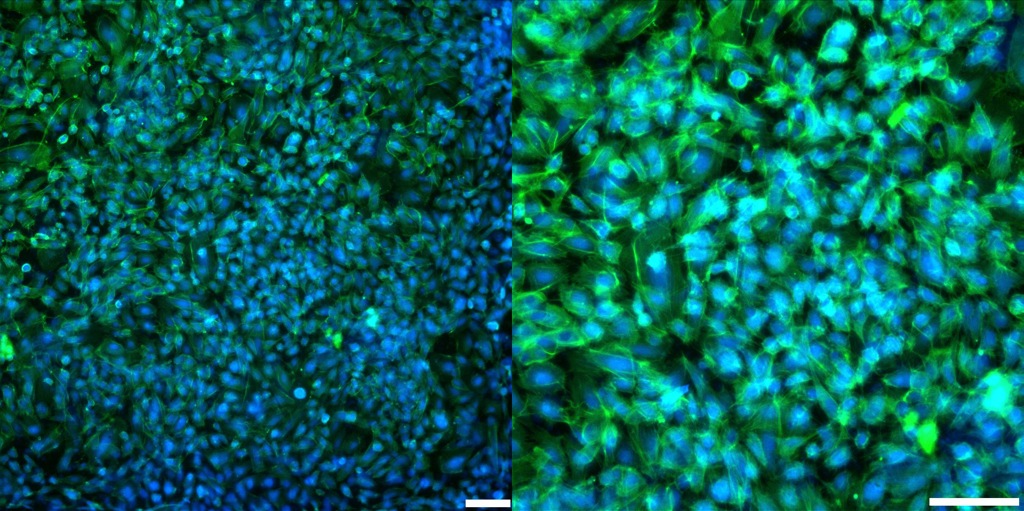
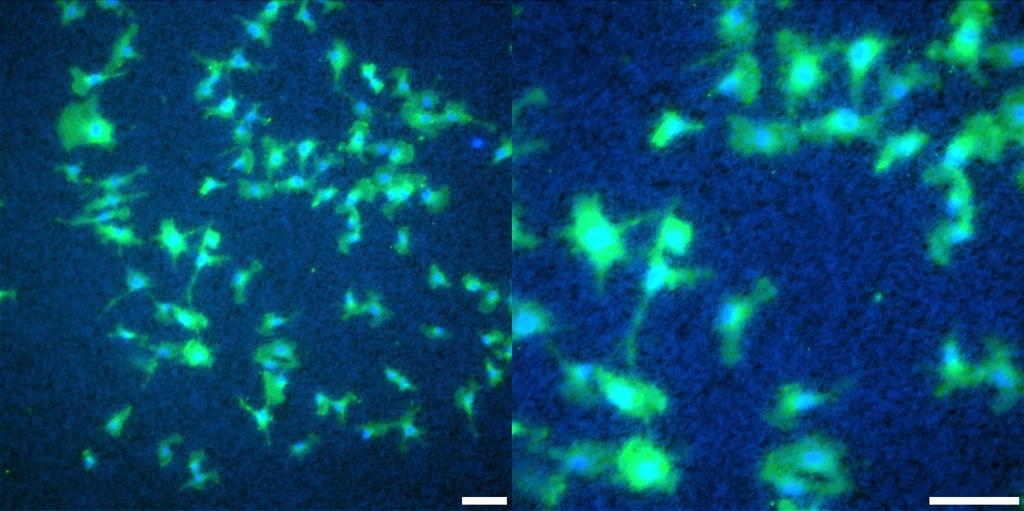
I recently performed the same staining protocol on other substrates: High Pore Density Microporous 300nm SiO2, Low Pore Density Microporus 300nm SiO2, Microporous SiN (~100nm), and Tissue Culture (TC) plastic. The cells were seeded at roughly 5*10^4 cells/cm^2, and were fixed, stained, & imaged 144 hours after seeding (as this data was originally meant for growth studies). Each surface was coated with 1% Geltrex (non-gel coating) before cells were seeded. Images were taken with either 90ms (DAPI) or 600ms (FITC) exposure time, and 0 EM Gain. Images were taken at 10x and 20x (all scale bars are 100µm).
Update!
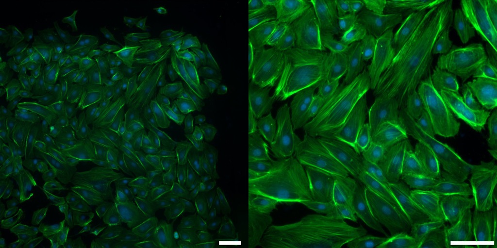
I also performed the fluorescence under the same coating and exposure/EM gain conditions for the 0.4µm pore size SiO2 membranes (both high and low pore density). The cells were fixed and imaged ~ 173 hours after seeding at 5*10^3 cells/cm^2. Scale bars are still 100µm.
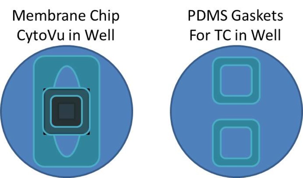
The setup of the two wells is as follows (comparing a CytoVu membrane and a TC plastic area):
The thin microporous SiO2 and track-etched (TE) images are from a previous trial (placed here for ease of comparison).
It seems that TC shows up most vibrantly and with highest resolution; this is understandable as all other substrates also had to pass through TC before the light could reach the cells regardless, and TC is highly optically transparent.
Microporous SiN, even though close to the thickness of the original SiO2 membranes (see previously referenced posts), has clearly visible pores even in the fluorescence. This does line up with the previous semi-quantitative study of fluorescent beads.
The two thicker SiO2 membranes seem more or less equivalent in terms of optical clarity and inability to see pores.
| Substrate: | 10x & 20x Combined Image |
| Thin 3.0µm Pore Size SiO2 | 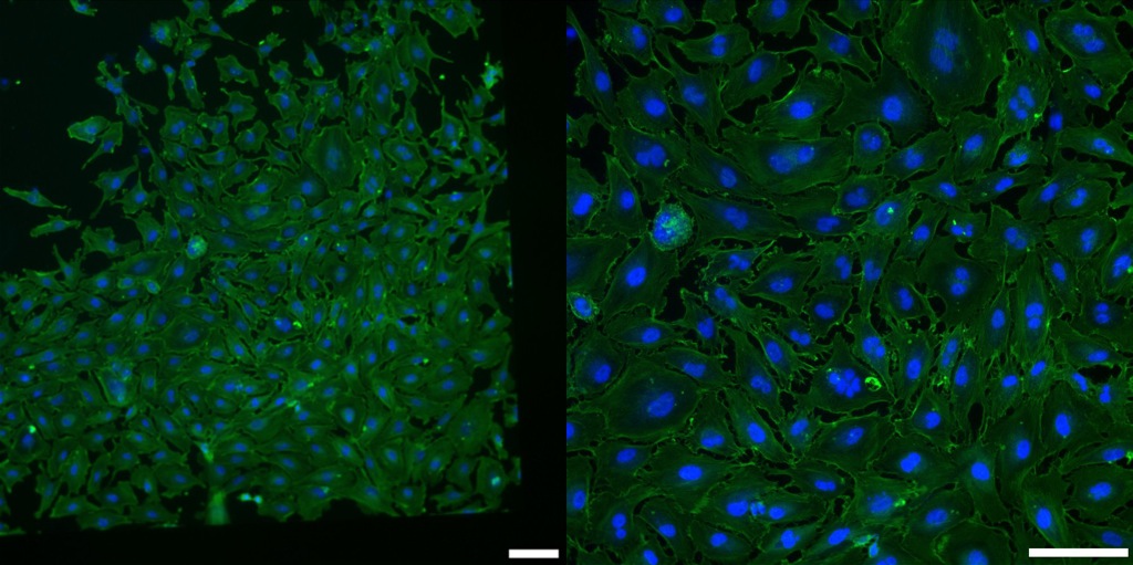 |
| Low Pore Density 3.0µm Pore Size 300nm SiO2 |
 |
| High Pore Density 3.0µm Pore Size 300nm SiO2 |
 |
| Low Pore Density 0.4µm Pore Size 300nm SiO2 |
 |
| High Pore Density 0.4µm Pore Size 300nm SiO2 |
 |
| Thin 3.0µm Pore Size SiN | 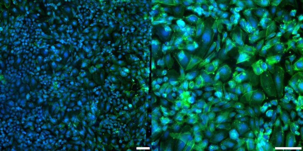 |
| TC Plastic | 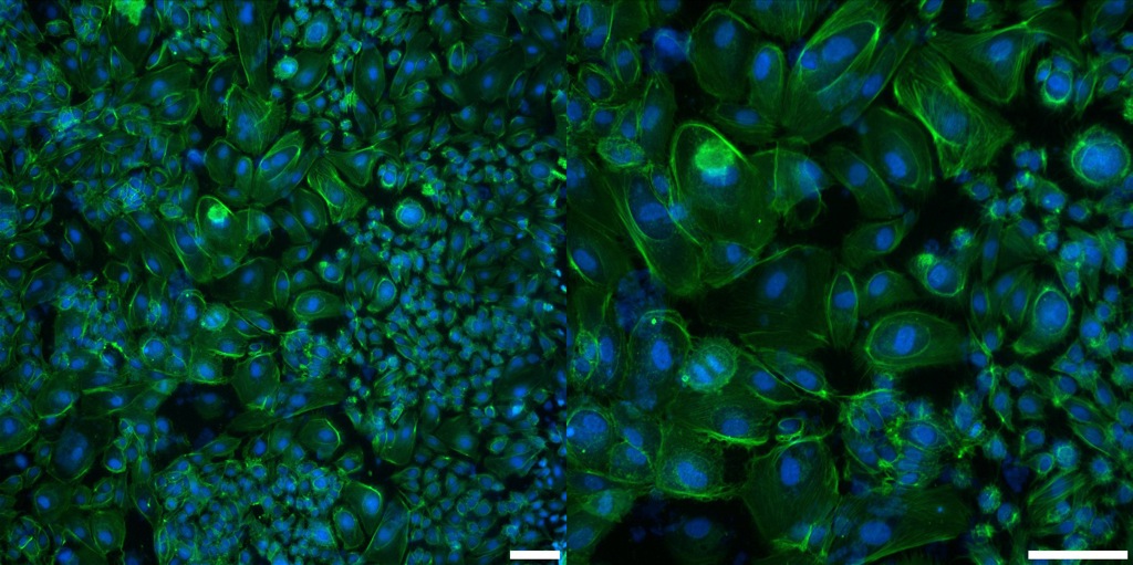 |
| TE CoStar 3.0µm Pore Size | 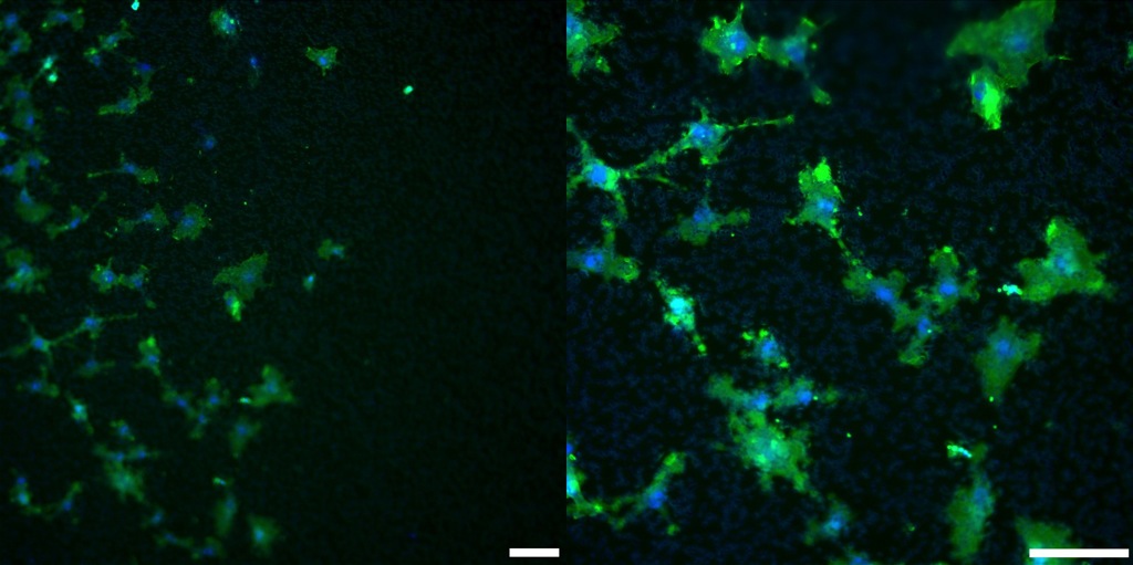 |
| TE CoStar 0.4µm Pore Size | 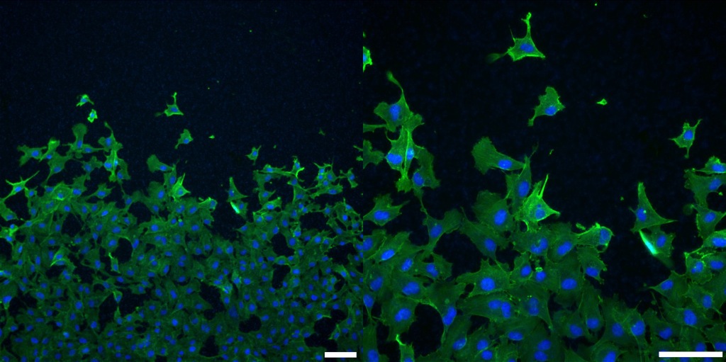 |
| TE Greiner 0.4µm Pore Size; ‘Translucent’ |
 |
| TE Greiner 0.4µm Pore Size; ‘Transparent’ |
 |
NRG: Update post to include track-etched images