Pharmacological Inhibition of Vacuole Formation
I’ve been trying to convince myself that the black holes seen in endothelial cells that we’ve been calling vacuoles are actually vacuoles. The first set of experiments involved staining cells with TAMRA, a fluid-phase marker which should get pinocytosed into vacuoles. Although there has been co-localization between ‘black spots’ in the calcein channel and red spots in the TAMRA channel, I haven’t seen all ‘vacuoles’ label red with TAMRA. I found a couple papers with experiments of pharmacological inhibition of vacuole formation, so I tried to apply these protocols to bEnd3 cells.
The drug is called bafilomycin A1. It’s an antibiotic that is also a specific inhibitor of vacuolar type H+ATPase (V-ATPase). V-ATPase is an enzyme that uses ATP to pump protons across different types of cell membranes (not just the plasma membrane). I seeded P18 bEnd3 cells at 50000 cells/cm2 on pnc-Si (SC502) and PET transwells. I let them grow for 1 day and then added bafilomycin (final concentrations 50, 100 & 200nM) to the media for 24 hours before staining with Live/Dead.
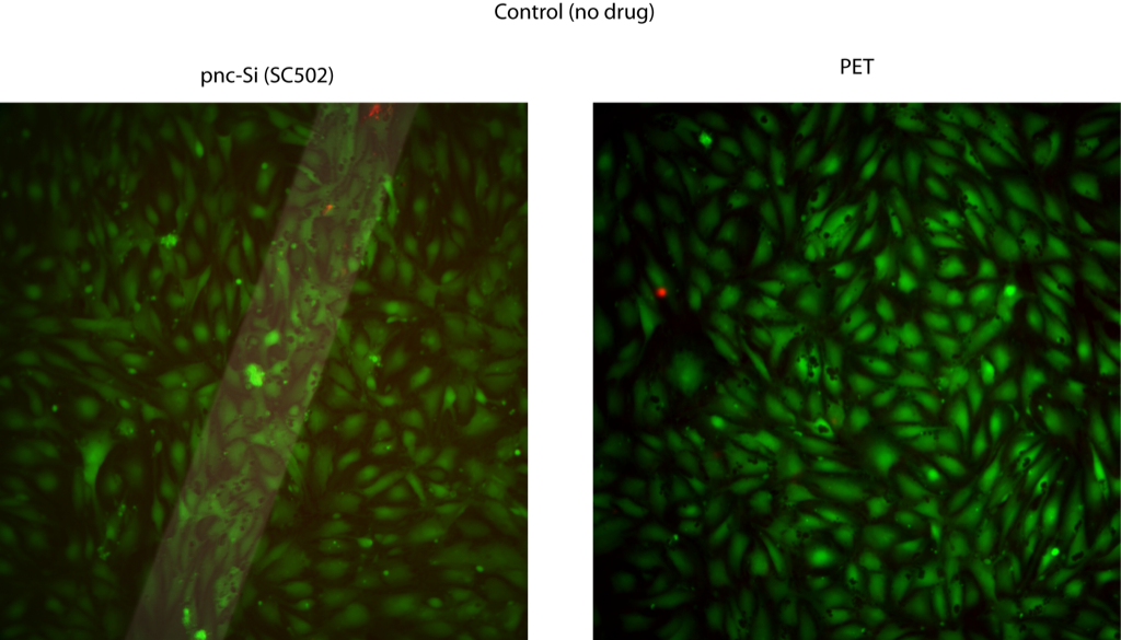
Control (untreated) Samples appear as they usually do:
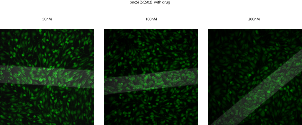
Vacuole formation was totally abolished pnc-Si transwells at all bafilomycin concentrations! Also, the cell morphology wasn’t too wacky and these drug concentrations didn’t kill the cells. It seems like there are a lot more cells over the membrane windows, especially in the 100nM and 200nM samples:
I saw the same results with PET transwells:
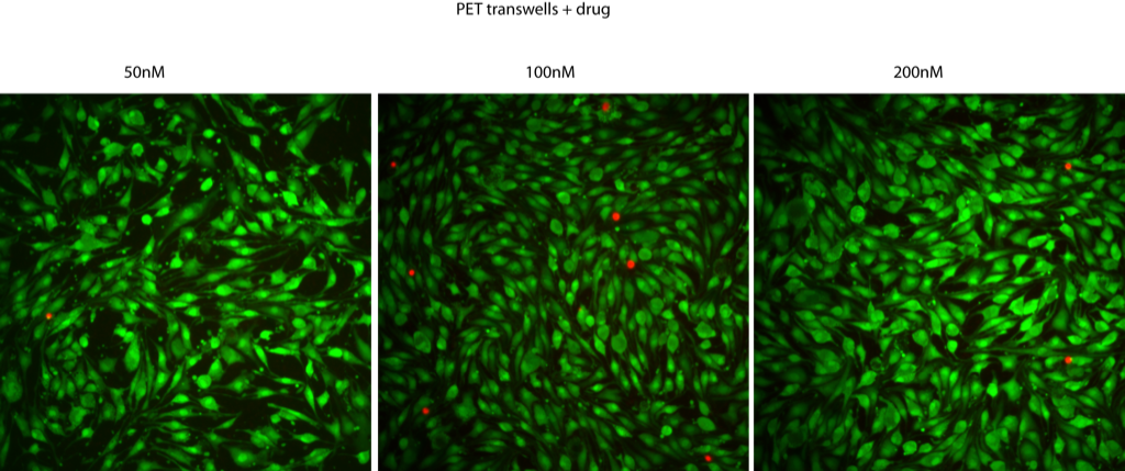
Based on these results, I feel confident that we are seeing vacuoles over membrane windows.