m-µSiM platform: TEER and Permeability for Open-Well iPSC-ECs and viability test for future flow module
Introduction
Tissue barriers regulate the absorption of nutrients, maintain homeostasis, and prevent toxins from entering. As previously introduced, trans-epithelial/endothelial electrical resistance (TEER) measurements offer a non-destructive and real-time method to quantitatively evaluate the integrity of tissue barriers. During the process, a small AC current (10 µA, 12.5 Hz) is applied across the barrier while the impedance measurements can be obtained over different time points to present the progression of the tightness of the barriers. When cell monolayer becomes confluent during incubation, pores of the membrane are covered by cells, preventing conductive ionic microchannels. And when the cells start forming tighter junctions, the impedance of the paracellular pathway for ionic current becomes higher – the tightness of the tissue junctions is proportional to the measured electrical resistance.
In this work, we try to map the TEER of a barrier to its permeability. As the TEER value of a barrier increases, the permeability of the barrier decreases. Therefore, TEER measurements can be used as a surrogate marker of the permeability of a barrier, and changes in TEER can be indicative of changes in barrier function.
We see the correlation between TEER and permeability – when the cell monolayer is healthy and the integrity of junction stays intact, the electrical resistance of the barrier is high, and the permeability is low. When the junctions are compromised due to damage, the permeability increases – leading to the free diffusion of solutes and ions across the cell layer, thus the electrical impedance of the barrier decreases. Therefore, TEER measurements can be used as a surrogate marker of the permeability of a barrier, and changes in TEER can be indicative of changes in barrier function.
When cells are exposed to shear stress, such as in a flow-driven culture system or a bioreactor, the TEER values may be different from their static culture counterparts. Shear stress can cause changes in the morphology and organization of the cell layer, which can result in changes in the tightness of the junctions between the cells. These changes can lead to an increase or decrease in TEER, depending on the specific conditions and cell type. For blood-brain barrier models, expose of mechanical stimuli may better represent the in vitro conditions.
Thus, in this work, we also introduce a magnetic latch-on lid for flow-based m-µSiM platform. We address some previous technical difficulties encountered and provide a future-proof design.
Theory
How TEER works
Cell layers can be modeled as an electrical analogy of a resistance and a capacitor. The resistance represents paracellular pathway related to cell-cell junctions and the transcellular pathway would be dominated by the capacitance of the cellular membrane/lipid layer. When we look into DC measurements or AC at relatively low frequencies (f -> 0Hz), the capacitance of the circuit (transcellular pathway) is considered fully charged and current cannot flow through the transcellular pathway. The tight junctions (paracellular pathway) in this scenario provide the major contribution to the impedance measured. Conversely, when the frequency of the voltage applied increases (f -> ∞), high frequency current can flow through the transcellular pathway, thus lowering the overall impedance of the cell monolayer.
Ideally, the resistance of paracellular pathway should be measured via DC electric field to quantify the tightness of junctions. However, since cell media are full of electrolytes, applying a DC voltage across the pair of electrodes may cause charging during the measurement period. This can lead to the pitting corrosion of electrodes, resulting in the passivity of the electrodes and releasing undesirable metal ions which can be detrimental to cells. Therefore, conventional TEER measuring methods use AC signal stimulations in the low-frequency regime, where the capacitance can still be considered as an open-circuit, to represent a DC measurement.
When the cell monolayer is not fully confluent across the porous membrane (m-µSiM), pores of the membrane, immersed in cell media, act as small ionic microchannels, allowing ionic current to flow through with a much smaller resistance.
Materials and Methods
Device fabrication
To create an accessible latch-on module compatible with the existing m-µSiM platform, we fabricated the TEER measurement lid and the lower housing for m-µSiM by laser-cutting acrylic sheets with thicknesses of 2 and 3.5 mm, respectively. The lower housing was bonded to a coverslip substrate using pressure-sensitive adhesive. Since commercial TEER readings with EVOM2 are highly dependent on the positions of the chopsticks electrodes positions (Srinivasan 2015), we design the TEER measurement lid with two electrodes fixed by 001 O-rings. The lid included two bonded acrylic layers to provide a stop of the O-ring to prevent slipping of the O-rings and thus of the electrodes. The lid fits on the m-uSiM based on magnetic latching between magnets embedded in the TEER module and lower housing as seen in Fig. 1.
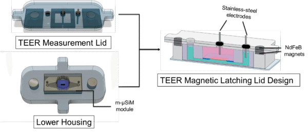
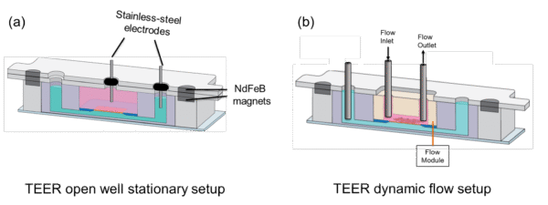
The m-µSiM platform allows us to perform cell culture with two formats: (1) open-well and (2) flow-driven. We have demonstrated the open-well measurement capability with this latch-on TEER lid. In the flow-driven culture format, we incorporate a PDMS-based flow module in the m-µSiM platform to drive flows using syringe tips and take TEER measurements periodically to evaluate the cell-cell junctions. Since our current electrode setup allows us to use stainless-steel syringe tips for measurements, this provides the opportunity to take TEER measurements without removing the flow module as shown in Fig. 2.
Cell culture
All reagents and steps were used based on the provided protocol by Engelhardt lab. µSiM devices with NPN membranes were treated with the coating solution (Collagen IV: 4 units, Fibronectin: 1 unit, Water: 5 units) for 3 hours in the incubator. iPSC-BMECs were split using Accutase (3 min) and seeded on each device at a density of 50,000 cells/cm2. The media was changed after 2 hrs of cell seeding. Immunostaining was conducted based on the protocol provided by McGrath lab. The cells were seeded at a density of 50,000 cells/cm2.
TEER measurements
By using a potentiostat (Reference 600, Gamry Instruments Inc.), we performed the impedance spectroscopy over a frequency sweep from 2.5 Hz to 250 Hz of iPSC-BMECs on m-µSiM under the open-well situation. Currently, we report our 12.5 Hz measurements to be consistent with the conventional EVOM TEER numbers in literature. We then took blank impedance measurements of the same setups without cells. The raw measurements subtracted by the blank impedance would provide the impedance the cell-cell junctions (paracellular pathway).
In this work, we also try to validate the flow-module with m-µSiM by performing a blank impedance measurement when compared to its open-well counterpart.
Results and Discussion
TEER lid on m-µSiM platform
The introduced magnetic latch-on approach allows users to seed cells and establish a monolayer in the open-well format. Once the cell monolayer is confluent, the user can latch-on the TEER measurement lid to characterize barrier resistance. Then, the TEER module can be removed to conduct the rest of the experiment in the open-well format. When taking TEER measurements, the Gamry Reference 600’s alligator clips can easily clamp onto the stainless-steel gauge.
In this work, we also introduce a flow-module insert design with addressed leakage issues we previously encountered.

We successfully drive flows from flow-module insert and back channel separated by the membrane. No leakage observed over 24 hours of observation period as shown in Fig. 4.

TEER measurements and permeability
We took measurements over 10 cell-seeded devices and 3 blank devices with m-µSiM setups.
Where indicates the impedance of cell-cell junctions, refers to the impedance measured using the potentiostat, and refers to the blank device impedance measurements. consists of the impedance of (m-µSiM + fibronectin/collagen-coating + cell media + electrodes). We use the 0-hr mark timepoint measurement of TEER as the blank impedance for each device.
To allow a more comparable value to literature, values are often reported by multiplying by the surface area of the porous membrane where cells are grown on. The porous window of the m-µSiM is 2 mm x 0.7 mm, yielding us the surface area of 0.014 cm2.
Raw impedance measurements for open-well vs dynamic flow setup yield some difference (15 vs 21 kΩ between open-well and flow setup) in blank impedance as shown in Fig. 5. COMSOL simulation yielded some difference 12.9 vs 16.5 kΩ (open-well vs flow setup).

We recorded cells on devices across 10 days of measurements. At day-11, we took the permeability of the devices. The results showed that the barriers with higher TEER measurements demonstrated lower permeability as seen in Fig. 6.

Conclusion and Future Work
We develop a latch-on TEER measurement lids compatible with the existing m-µSiM platform (Mehran, 2022). The m-µSiM platform allows us to perform cell culture with two formats: (1) open-well and (2) flow-driven. We have demonstrated the open-well measurement capability with this latch-on TEER lid. In the flow-driven culture format, we incorporate a PDMS-based flow module in the m-µSiM platform to drive flows using syringe tips and take TEER measurements periodically to evaluate the cell-cell junctions. Since our current electrode setup allows us to use stainless-steel syringe tips for measurements, this provides the opportunity to take TEER measurements without removing the flow module for the microfluidic flow driving format.
Future work would feature co-cultures of endothelial cells with pericytes or astrocytes for valuable insights when creating in vitro models of tissue barriers, as endothelial cells are responsible for forming junctions, both pericytes and astrocytes seem to participate in the formation.