Microengineered three-dimensional collagen fiber landscapes with independently tunable anisotropy and directionality
The extracellular matrix in vivo is composed of fibers arranged with varying degrees of alignment in different directions. The extent of alignment (anisotropy) and the directionality of fibers are an indicator of breast cancer metastasis, influence T-cell motility, and guide endothelial cell alignment. Similar to the existence of chemical gradients and stiffness gradients, the ECM is also characterized by heterogeneity (or gradients) in the collagen fiber microstructure. ECM fibers are observed to transition to an aligned organization at a tumor boundary, thus directing invasive tumor cells away from the tumor site. The degree of ECM alignment has also been observed to increase with increasing tumor potency. However, current techniques to control collagen fiber orientation fall short of recapitulating the spatial gradients in fiber alignment since they can only control alignment in a single direction.
This study aims to create gradients in collagen fiber alignment, exploiting a microfluidic channel with varying cross-sectional areas to generate varying degrees of extensional strain in the collagen prepolymer solution, resulting in collagen fiber alignment that is directly related to the applied strain rate.
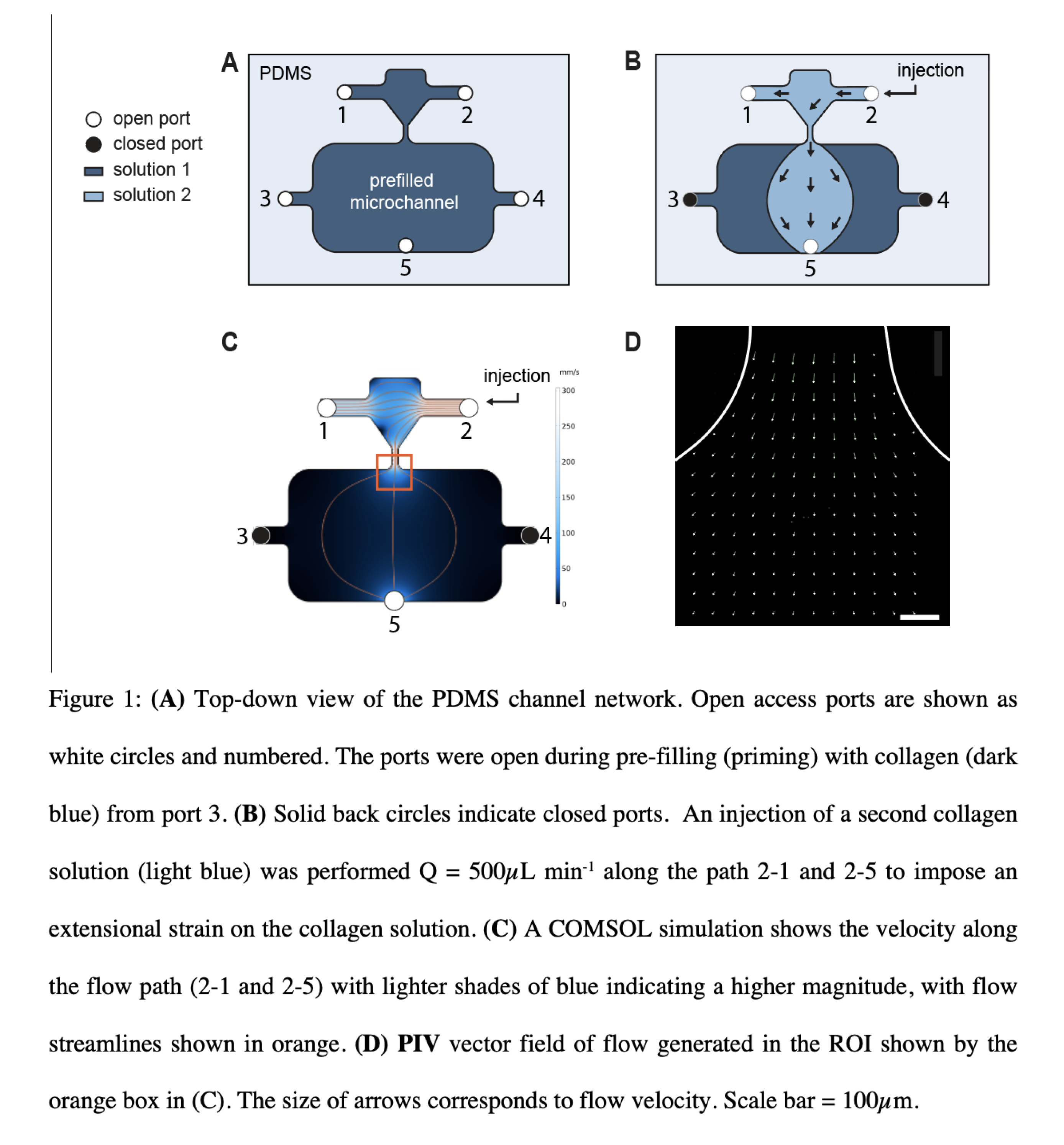
We designed a microfluidic channel with 5 fluid access ports and a variable cross-sectional area along the flow paths (figure 1). The dimensions of the channel are shown. The channel was made out of PDMS and reversibly placed on a glass coverslip. For initial experiments, we used a 2 -stage injection process to fill the channel. All ports were open to atmospheric pressure for the prefill, and neutralized collagen (2.5mg/ml) was preloaded into the channel from port 3 at a slow flow rate (~4µl/min). After prefilling, ports 3, 5 were closed using tape, and a neutralized collagen solution was injected into the channel from port 2 at a high flow rate of 500µl/min. The injection process lasted for 5 seconds and resulted in a peak strain rate of ~130/s at the constriction in the path from port 2 – 4. The device was placed in the incubator for 2 hours before imaging.

As seen in figure 2, imaging was carried out in 2 regions – from port 2-1 and at the constriction exit from port 2-4. Collagen alignment at the inlet region was found to be more or less constant with a coefficient of anisotropy ranging from 0.6 – 0.69. The directionality of collagen fibers from port 2 to the constriction was found to change gradually from 0˚ at port 2 to 75˚ towards the constriction. Therefore, this platform offered control over collagen fiber orientation while maintaining fiber anisotropy. In figure 2B, the region immediately after the constriction was imaged. Fluid exiting the constriction undergoes a rapid drop in strain rate, and a corresponding decrease in the coefficient of fiber anisotropy was observed in the region. To the best of our knowledge, this is the first demonstration of creating gradients in fiber anisotropy using microfluidic systems.

The 2-stage injection process was also used to make material interfaces. To test the ability to make interfaces, collagen was prefilled into the channel with all ports open, from port 3. Immediately after, ports 3, 5 were closed, and a blend of neutralized collagen with 5 wt% HA and 0.1% fluorescent beads was injected from port 2 at a flow rate of 500µl/min. The injection process lasted for 5 seconds. Due to a purely laminar flow in the channel at the specified flow rates, we predicted that the 2 fluid types would undergo minimal mixing. In figure 3B, we can observe that the beads are localized in the region expected to be occupied by the collagen HA mixture. The localization of fluorescent beads suggests the presence of a material interface in the region. Fiber anisotropy was also observed in the collagen HA mixture. Multiple fluid access ports can be opened or closed to direct fluid flow and thus be exploited to create custom interfaces between 3D materials.

Most microfluidic channels with 3D ECM gels pose practical issues towards introducing cells due to limited access to the ECM gel through side channels and ports. To enable easy cell culture on the microengineered collagen landscapes, we implemented the ability to lift off the PDMS microchannel after collagen gel formation and expose the landscape for direct access. The PDMS channels were coated with 4% BSA for 4 hours to passivate the surface and prevent collagen adhesion, while the glass coverslips were coated with a thin layer of poly(octadecene-alt-maleic anhydride) (POMA) to enable covalent attachment of collagen. After gel formation, the PDMS channel could be lifted off using tweezers, shown in figure 4A. Figure 4b shows representative images of the same region of collagen fibers before and after channel lift-off, suggesting that the lift-off process had minimal effect on the fiber alignment. Further, an analysis of the change in CoA across 50 ROIs showed that collagen fiber CoA was minimally affected by lift-off. By implementing channel lift off, we can eliminate the need for delivery ports for media delivery and make experimental processes such as immunostaining and RNA extraction easier.

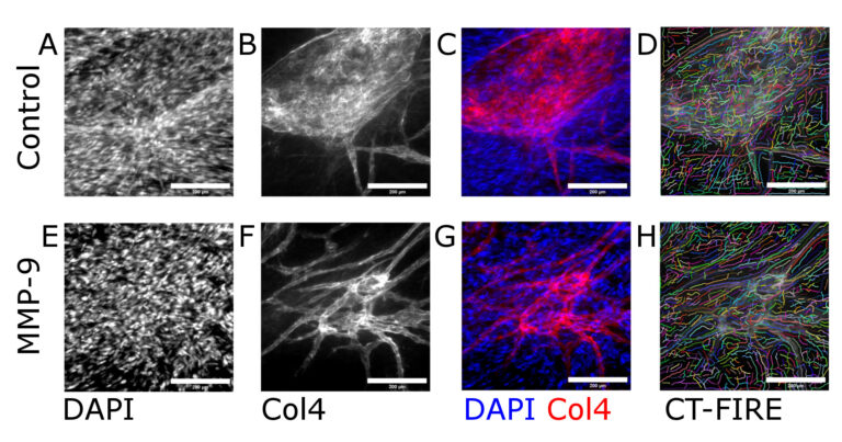
To test the response of cells to the microengineered gels, MDA-MB-231 cells were cultured on the platform. After 24 hours, the cells were found to align in the direction of collagen fibers (figure 5A), and they displayed an elongated morphology. The aspect ratio of cells on aligned collagen was significantly higher than the aspect ratio of cells on randomly oriented collagen fiber control (Figure 5B). Similar results were observed with HUVECs, which were cultured as a sheet on the aligned collagen. Actin fiber alignment of HUVECS on aligned collagen displayed a narrower spread as compared to the control sample (Figure 5C)
In conclusion, this work shows:
- The use of microfluidics to achieve gradients in collagen fiber anisotropy and directionality
- Fabrication of 3D material interfaces in a fluidic network
- Easy channel lift-off for direct access to microengineered collagen
- The response of MDA-MB-231 cells and HUVECs to aligned collagen