The effect of Exosomes on Wound Healing
Hello Everyone,
The goal of these experiments was to study the effect of exosomes on wound healing. We wanted to study, first importance of exosomes for migration of FBs and eventually closure (healing) of the wound and second effect of exosomes derived from ADSCs on FBs migration. In order to test our hypothesis, we designed 6 sets of conditions including:
- No wound: with fresh media as the baseline: (Media Nutritions + Serum Exosomes)
- DMEM + Exosome Free FBS: (Media Nutritions)
- DMEM + FBS: (Media Nutritions + Serum Exosomes)
- 1 Day ADSC Conditioned Media: (Media Nutritions + Serum Exosomes +1 Day ADSC Exosomes)
- 3 Day ADSC Conditioned Media: (Media Nutritions + Serum Exosomes +3 Days ADSC Exosomes)
- 1 Day FBS Conditioned Media: (Media Nutritions + Serum Exosomes +1 Day FB Exosomes)
We hypothesized that cells need both Media Nutritions and Exosomes for migration. In cases of conditioned media, the media nutitions and serum exosomes have been consumed by the cells partly or completely, but certainly, the amount of the nutritions and serum exosomes after 3 days is less than after 1 day. It’s important to note that, making a fair conclusion is not possible with these conditions since more than only one factor have changed between conditions. Ideally, exosomes need to be isolated from each of this conditions and be added to the DMEM (Media Nutritions) to study the effect of exosomes. But, this experiment is still useful for studying this set of conditions. Concentration of particles were measured and analyzed by Nanosight.

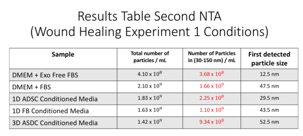
As you can see here, DMEM + FBS has more exosome size particles showing the presence of exosomes in the Serum. 1 day ADSC sample has less particles compared to both DMEM + Exo free FBS and DMEM + FBS samples meaning that most likely, the media nutritions and serum exosomes have been consumed by the cells (partly or completely) and some exosome size particles have been produced by ADSCs. 1 day FB sample shows a high concentration possibly meaning high production of exosomes. In addition, the 3 day ADSC conditioned media is showing higher concentration compared to 1 day conditioned media possibly meaning more exosomes have been secreted over time.
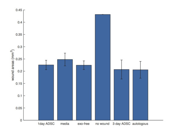
Fibroblast cells were grown to a confluence layer on a 96 well plate and they were scratched using a pipette tip (P10) for Wound formation. 4 wells were used for each set of conditions, and 3 different region of interests were imaged (2 on the wound and 1 on without wound region). The time lapse images were analyzed by Henry’s migration Matlab Code. As the first step, we measured the wound area for different conditions. it’s important to note that the wound area for the no wound condition is actually zero.
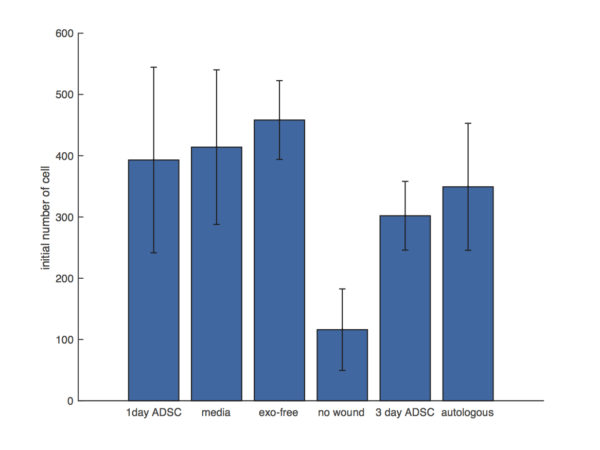
second, we wanted to see what is the initial number of cells for each experiment condition;
Furthermore, the number of cells appeared in the wound region was monitored for different conditions. And the percent cell density recovery was defined as the ratio of number of cells appeared in the wound region to the the number of initial cells (Before wound) in the wound area. The ratio shows us the recovery rate of the wound with 100% meaning full recovery.

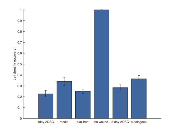
As you can see from the results the autologous condition (1 day FB conditioned media) shows the best cell density recovery following by the fresh media (DMEM + FBS). Surprisingly, exosome free condition and 1 day ADSC condition do not show a significant difference that might be due to the fact that not all the exosomes are actually depleted in exosome free FBS sample.