Cellular Adhesion: Bare NPN v PEG NPN
In 5.4 mm single slot chips, sheep blood was flowed at 3 µL/min though the 3 mm x 1 mm channels with PBS flowed at the same rate but in the opposite direction for four hours. The chips were pre-wetted with PBS and rinsed following flow. The membrane surfaces were fixed with 4% paraformaldehyde for >10 minutes and rinsed again.
One set of chips (3) were untreated, BARE, while the other group (3) were coated in Dr. Shestopalov’s lab (by Xunzhi) with PEG. Using an optical microscope, ImageJ, and the pore processing matlab tool, the percent of area covered by cellular material was measured.
It is evident from the 5x images that the PEG was successful in reducing cellular adhesion. Higher mag images (50X) were used with the pore processing software to determine the ‘porosity’, which I am using to measure not pores but debris so the percentage the software reports is the percent of the surface covered by cellular material.
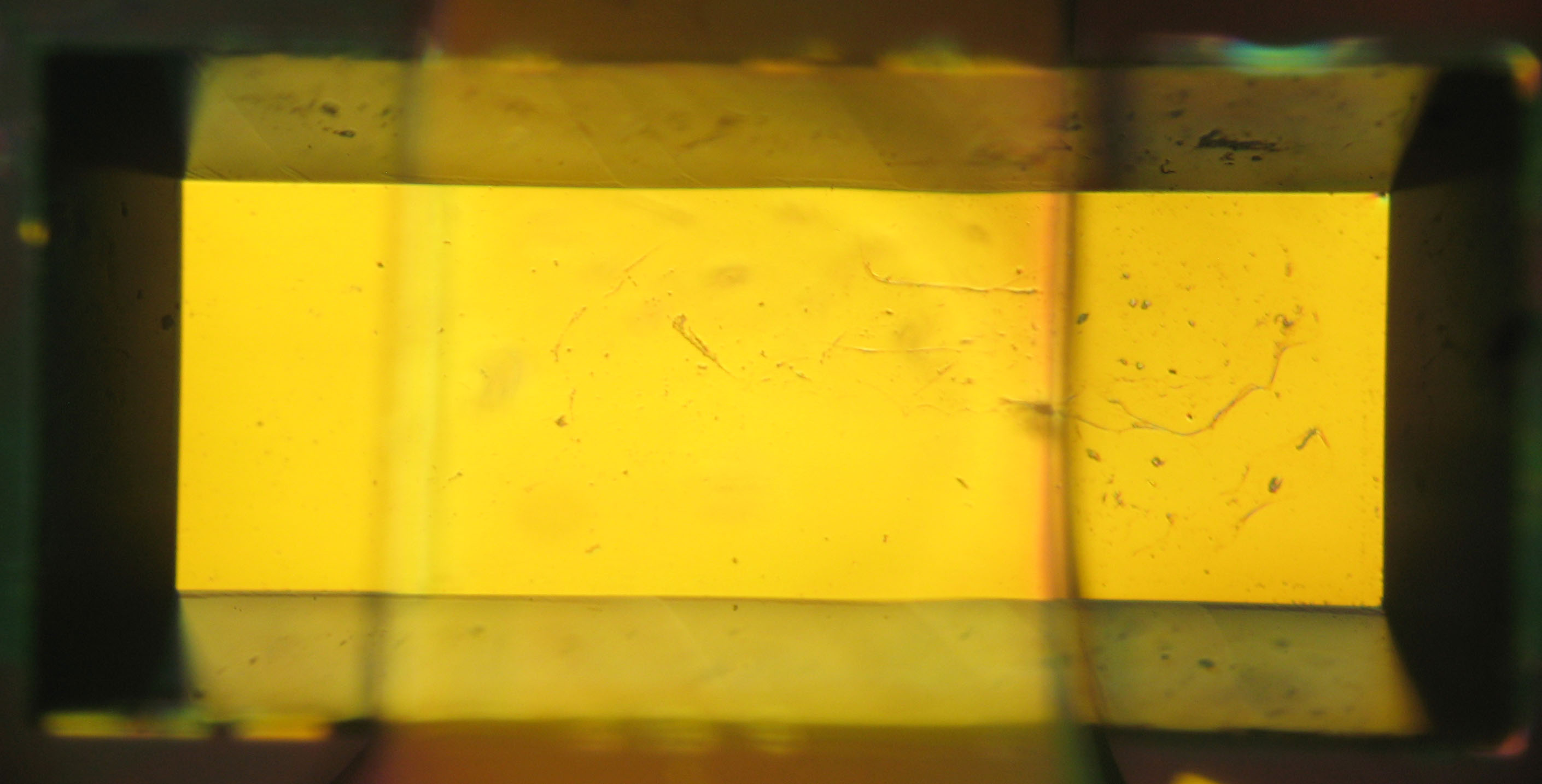
Bare membrane vs PEG membrane at 5X

Bare membrane vs PEG membrane at 50X, near one end of the channels
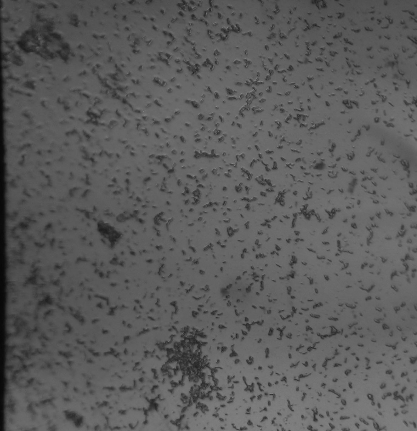
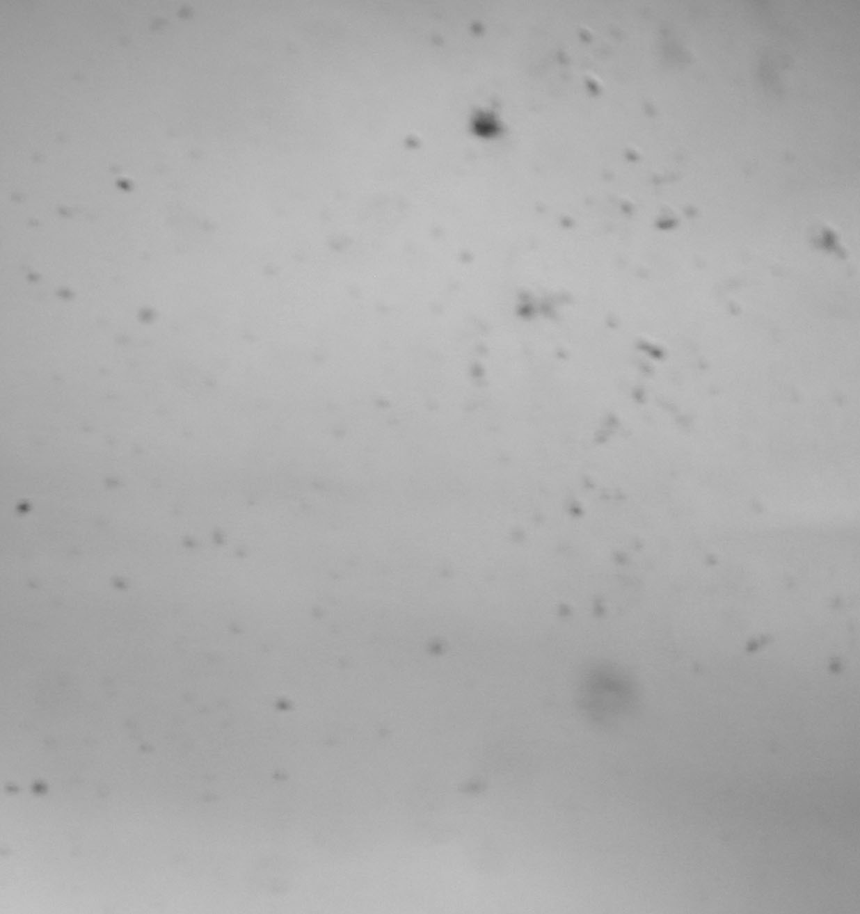
Similar to the effect seen in the small animal studies where the contamination was heaviest at the edges of the membrane, the untreated membrane had a higher percentage of fouling on both ends of the channel (15% vs 7%). The PEG treated membranes had little fouling anywhere and in fact has less fouling at the ends of the channel (0.4% vs o.25%).
This is good news for protein binding and the next step is to plan and schedule one more round of the small animal study.
