Pore processing of one backside image of 75 nm thick membrane
Here is a better, more thoughtful, attempt at pore processing of a single image of the backside of a 75-nm thick membrane (four slot).
One thing you will note that could be different from the analysis of the frontside of the membranes: The small openings that appear quite sharply defined here could be missing from the frontside images, looking either like pits or perhaps larger pores. This, if true, lowers the measured average pore size from previously reported values. I suppose it would be possible to image the same membrane from both sides at the same location if you registered the image from the corner of the membrane. Then a direct comparison of the two methods could be made. Just a thought. I do realize that this image, at 100,000 x, is highly magnified and therefore has a small number of pores. My images were taken to see the surface and not intended for pore processing so I have high, medium, and low mag images for future processing. In the future I can save more images at a more optimal mag for pore processing if this mag proves to be too large.
JingKai “JK” Zhang will be helping process more images this summer so I wanted to hone the process to make sure the results are sensible and inline with previous work on the frontside images.
The original image on the left and the inverted image on the right. The software needs white holes and a dark field.
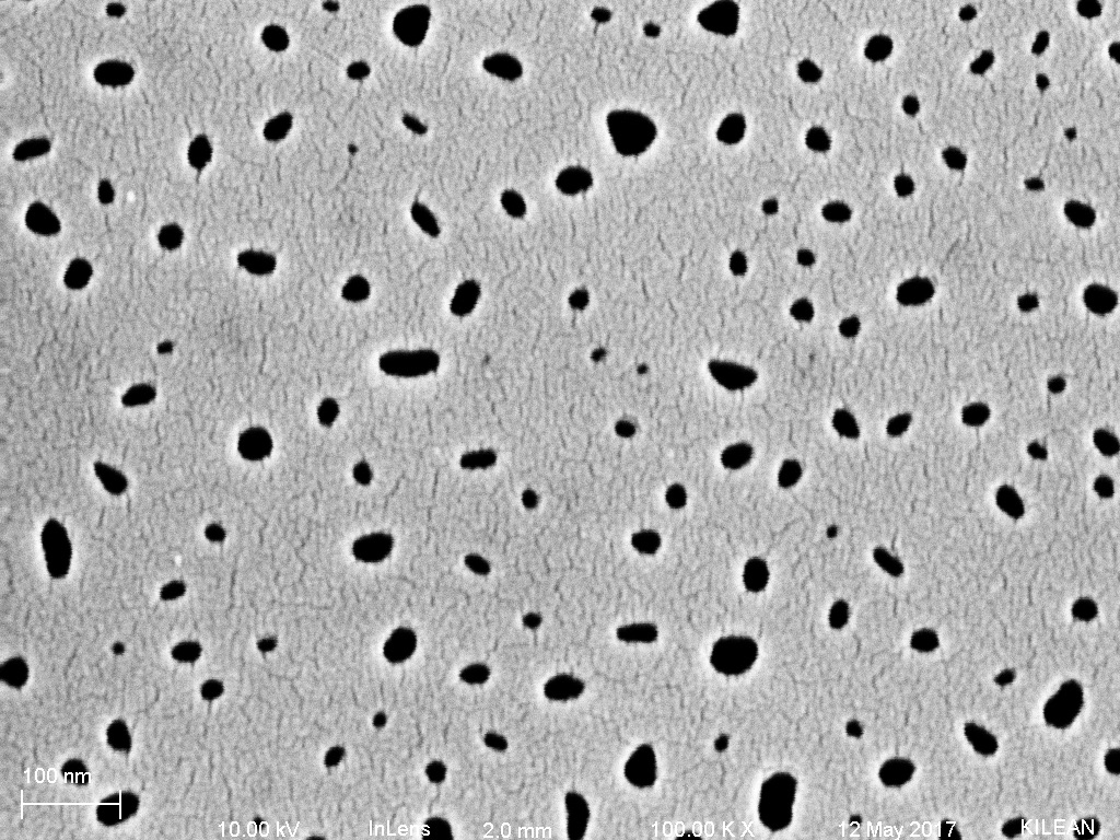
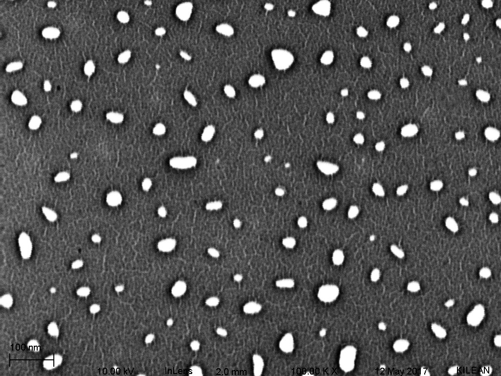
The next two images are the results from the pore processing itself.


And the histograms. Mean pore:
Diameter: 23.6
Major Axis: 28.7
Minor Axis: 19.8

